Download
1 / 62
1.86k likes | 5.95k Vues
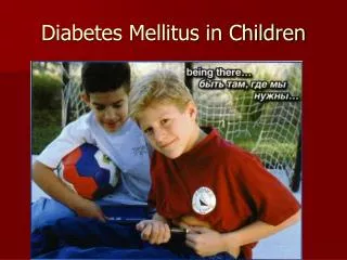
Diabetes mellitus in children. By Henry Cummings MBBS, FMCPaed Delsuth , Oghara. Pre-test. Dm is the commonest endocrine disorder in children Only type 1 dm occurs in children TIDm is a non progressive low-insulin catabolic state

E N D
Diabetes mellitus in children By Henry Cummings MBBS, FMCPaed Delsuth, Oghara
Pre-test • Dm is the commonest endocrine disorder in children • Only type 1 dm occurs in children • TIDm is a non progressive low-insulin catabolic state • Exogenous insulin replacement remains the only form of replacement therapy • In managing DKA, always add kcl to the initial rehydrating fluid.
Childhood Diabetes mellitus • Definition • Types • Patho physiology • Clinical features • Modalities of management • Complications, short term, long term • Recent advancements • Conclusion
DEFINITION BY WHO: • DM is a metabolic disorder of multiple aetiologiescharacterised by chronic disturbances of carbohydrate, fat & protein metabolism, resulting from defects in insulin secretion, insulin action or both. • DM is the commonest endocrine disease in childhood & adolescence.
MAGNITUDE OF THE PROBLEM • Overall incidence 1 to 2 per 1000 school age children. • Estimated prevalence of childhood DM in 2003 were: • 430,000 globally • 250,000 live developing countries. • 63,000 live in 58 poorest countries(least developed countries).
TYPES OF DM Type 1 DM(IDDM) • Beta cell autoimmune destruction • Absolute insulin deficiency • Requires insulin for survival • Accounts for over 90% of childhood DM • Peak age incidence 10-12 years • Slight male predominance • Prone to ketosis
Type 2 DM (NIDDM) • Insulin resistance with relative deficiency OR • Secretory defect with or without resistance • Does not require insulin for survival • Strong genetic component • Not prone to ketosis • Acanthosisnigrans may be present • No islet cell antibodies • Associated with overweight teenagers
Maturity onset diabetes of the youth (MODY) Early onset of dominantly inherited type 2 DM Non- obese children No islet cell antibodies Family history in several generation Identified genetic mutations e.g mutations of glucokinase or hepatic nuclear factor 1 & 2 genes Non- ketotic Two (or at least one)family member diagnosed before age of 25 years
Neonatal Diabetes • hyperglycemia requiring insulin in the first 3months of life • Rare condition 1:400,000 • Associated with IUGR • 50% of cases are transient • Associated with paternal isodomy& other imprinting defects of Chr 6 • In transient NND permanent DM may appear later in life
Secondary diabetes mellitus • May be associated with cystic fibrosis, hemochromatosis, drugs such as L-Asparaginase
Criteria for diagnosis • Symptoms of DM and casual plasma glucose conc > 11.1mmol/L(200mg/dl) (10 for venous) • FPG > 7.0mmol/L(126mg/dl) (6.3 for venous and cap) • 2hr post load of glucose >11.1mmol/L during an OGTT
blood sugars • Normal RPG: 70 – 140mg/dl • Normal FBS: 70 – 100mg/dl • Hypoglycemia: • Mild 40 – 70mg/dl • Severe 40mg/dl • Neonatal <50mg/dl
Etiology of T1Diabetes • Environmental • Factors • Cow’s milk? • Viruses ? • Nitrates? • Genetic • Susceptibility • DM1: HLADR3,DR4↑ • Protective DRB1,DQB1↓ • DM2 Autoimmunity & Insulitis Destruction of pancreatic βcells
Pathophysiology Insulin • essential to process CHO, fat, protein • It blood glucose levels by glucose uptake into muscle cells and fat cells • stimulates glycogenesis • inhibits glycogenolysis • the breakdown of fat to triglycerides, free fatty acids, and ketones. (lipolysis)
Pathophysiology Lack of Insulin • glucose oxidation in muscle & fat cells • Proteolysis & amino acid release • Glycogenolysis & gluconeogenesis • all result in Hyperglycaemia • Glucose intake still continues
Pathophysiology • The kidneys cannot reabsorb the excess glucose load, causing glycosuria, osmotic diuresis(polyuria), thirst, and dehydration. • Untreated pt excrete high glucose load causing polyphagia • Increased fat and protein breakdown leads to ketone production and weight loss.
Pathophysiologycontd • Acidosis result from ketosis • Acidosis leads to renal excretion of K+ and Po4 • Na+ loss is due to osmotic diuresis & vomiting • Hypokalemia is due to vomiting, osmotic diuresis & hyperaldosteronism • Coma likely due to ketosis, acidosis, dehydration & hyperosmolality
Absolute insulin deficiency OR Stress, infection or insufficient insulin intake Counter-regulatory hormones: Glucagon, Cortisol, Catecholamines, GH Lipolysis • Glucose utilization Proteolysis Protein synthesis Glycogenolysis Gluconeogenic substrates Gluconeogenesis FFA to liver Ketogenesis Hyperglycemia Alkali reserve Glucosuria (osmotic diuresis) Acidosis Loss of water and electrolytes Dehydration Lactate Hyperosmolarity Impaired renal function
Phases of T1DM • Preclinical diabetes • Presentation of DM • Partial remission or honeymoon phase • Chronic phase of lifelong dependency on administered insulin
Preclinical DM • Occurs months to years preceding the clinical presentation of T1DM • Antibodies can be detected as markers of beta cell auto immunity:- GAD, IA,ICA, IA2 etc • IVGTT
T1DM: a slowly progressive T-cell mediated autoimmune illness “Silent” Cell Loss Inciting Event(s) Diabetes Diabetes Onset I II III • cell Mass?? Is cell loss exclusively immune mediated? Time (years) Genetic susceptibility 100% Islet Cell Mass 50% 0%
Clinical presentation • Vary from non-emergency px( polydipsia, polyuria, weight loss, enuresis) to severe dehydration, shock and DKA • Onset may be acute, precipitated by an acute illness, or more chronic and insidious over weeks or even months.
Lab Investigations • Glucose levels • E & U • Ketones • C peptides • Islets cell antibodies
Other Lab Investigations • Lipids • Microalbumin • Thyroid fxn test • Hb A1c • fructosamine
Modalities of management • requires Multidisciplinary team • Insulin therapy • Diabetic education • Exercise • Diet • Psychological care • Monitoring • Others:- sick day mx, adjusting to school
Insulin therapy • Exogenous insulin replacement remains the only form of replacement therapy
The Basal/bolus Insulin Concept • Basal Insulin - Suppresses glucose production btw meals and overnight • 40% to 50% of daily needs • Bolus Insulin (meal time) • Limits hyperglycaemia after meals • Immediate rise and sharp peak at 1 hour • 50% -60% total daily insulin requirement for meals
Basal/bolus therapy regimens Intensive management: • MDI – Multiple Dose Insulin • Once-daily IA or LA insulin usually given at night and 3-ce daily SA or VA before each meal • Mixed preps e.g. Mixtard, humulin 70/30 • CSII – Continuous Subcutaneous Insulin Infusion (Insulin pump therapy)
INSULIN THERAPY contd. • Twice daily regimen: • Split dose regimen (2/3 morning, 1/3 evening; 2/3 intermediate acting, 1/3 soluble). Aim at maintaining blood glucose within 80-150mg/dl, with the occurrence of as few hypoglycaemic episodes as possible.
60 40 Insulin 20 0 Twice a day insulin Endogenous insulin Soluble insulin Intermediate-acting insulin Breakfast Lunch Supper
DIET • Complex carbohydrates (CBHs) are preferred to simple refined CBHs. • Dietary regimen should be adjusted according to convenience of the family and school timings to ensure better compliance. • Total CBH content of the meal &snacks should be kept constant.
EXERCISE • Encourage regular exercise. • Insulin requirement may be lower, metabolic control improved and self-esteem & body image better in physically fit child.
SELF- CARE EDUCATION • Should include nature of illness, acute & chronic complications, insulin action, duration and timing, injection techniques, nutrition information, self blood glucose monitoring and urine ketone checks. • Education must be appropriate to child’s age & family educational background.
Monitoring • Monitoring of growth & development: the use of percentile charts is a crucial element in the care of children & adolescents with DM • Poor gain of height & weight, hepatomegaly and delayed puberty might be seen in children with persistently poorly controlled DM
Monitoring • HbA1c at least twice a year • Screening for long term complications
Partial remission (honeymoon) phase • Defined as when the patient requires < 0.5units of insulin/kg/day and has an HbA1c <7% • Due to partial recovery of the damaged beta cells • Commences within days or weeks of start of insulin therapy and may last for weeks to months • Ketoacidosis at presentation & young age reduces the likelihood of a remission phase
Complications • comprised of 3 major categories: 1. acute complications - reflect the difficulties of maintaining a balance between insulin therapy, dietary intake, and exercise. 2. long-term complications 3. complications caused by associated autoimmune diseases
ACUTE COMPLICATIONS • Hypoglycaemia • Diabetic ketoacdosis • Infections
HYPOGLYCAEMIA • Defined as blood glucose level < 60mg/dl (3.3mmol/L). For preschool children, values below 70mg/dl (3.9mmol/L) should be a cause for concern. • Severe episodes occur 10-25% of pts per year • Commonest acute complication of Type 1 DM
WHY WORRY ABOUT HYPOGLYCAEMIA? Recurrent severe hypoglycaemia can lead to: • Hypoglycaemia unawareness(25% of DM pts) • Epilepsy • Learning difficulty • Death (accounts for 4% of deaths in DM as a result of unintentional trauma).
Diabetic ketoacidosis • creates a life-threatening medical emergency. • is the most important cause of mortality and severe morbidity in children with diabetes, particularly at the time of first diagnosis. • Early recognition and careful management are essential if death and disability are to be avoided.
Diagnosis • 3 cardinal features: • Hyperglycemia - >200mg/dl(11.1mmol/l) • Ketonuria >5mmol/l, ketonemia • Venous ph<7.3 or metabolic acidosis < 15mmol/l • Clinical features • Severe dehydration, shock • Frequent vomitng • Polyuria despite dehydration • Weight loss in spite of good intake • Acetone breath – (Kussmaul respiration) deep and rapid • Altered sensorium • Signs of raised intracranial pressure – bradycardia, HT, anisocoria
RISK FACTORS • AS INITIAL PRESENTATION: • Young age: < 5 years • Low socioeconomic background • IN ESTABLISHED TYPE 1 DM: • Higher HbA1c • Adolescents, particularly females • Psychiatric disorders • Longer duration of diabetes
severity • mild = pH < 7.30 or bicarb < 15 mmol/L • moderate = pH < 7.20 or bicarb < 10 mmol/L • severe = pH < 7.10 or bicarb < 5 mmol/L
Goals of therapy • Correct dehydration • Correct acidosis and reverse ketosis • Restore blood glucose to near normal • Avoid complications of therapy • Identify and treat any precipitating event
Emergency assessment • Brief history to find cause • Weigh the child • Assess degree of dehydration • Assess level of consciousness(glasgow coma scale) • Biochemical assessment
Biochemical assessment • Blood samples: • Plasma glucose • E&U, Cr, Ca, PO4, Mg • HbA1c • Venous pH • pCO2 • Hb • FBC( Leucocytosis could exist without infection due to stress) • Beta hydroxybutyrate
Biochemical assessment • Urine sample • Urinalysis for ketones (acetoacetate) Others include culture samples:- (blood , urine, throat) ECG:- k status
Supportive therapy • Secure airway • A peripheral IV catheter should be placed in for convenient and repetitive sampling • Cardiac monitor • Give oxygen to pts with severe circulatory impairment or shock • Give anitbiotics to febrile patients after cultures have been taken
FLUID THERAPY • 1st hr: 10 – 20ml/kg 0.9% Nacl with insulin infusion at 0.05 – 0.1 U/kg/hr • 2nd hr & subsequent hrs: 0.45% Nacl plus continuous insulin drip + 20mEq/L K+ • 5% dextrose if blood glucose <250mg/dl (14mmol/L) • IV Rate= 85ml/kg +(maintenance minus bolus) divided by 23