V7: Cell differentiation
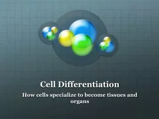
Cell differentiation in complex genomes leads to a variety of cell types. Transcriptional programs and epigenetic modifications play vital roles in lineage determination during development. Explore how germ cells are produced from zygotes and the differentiation of somatic cells from gametes in humans and other organisms.

V7: Cell differentiation
E N D
Presentation Transcript
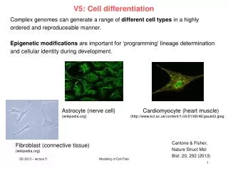
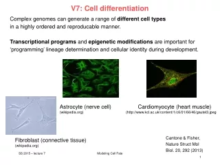
V7: Cell differentiation Complex genomes can generate a range of different cell types in a highly ordered and reproducable manner. Transcriptional programs and epigenetic modifications are important for ‘programming’ lineage determination and cellular identity during development. Astrocyte (nerve cell) Cardiomyocyte (heart muscle) (wikipedia.org) (http://www.kcl.ac.uk/content/1/c6/01/66/46/gautel3.jpeg Cantone & Fisher, Nature Struct Mol Biol. 20, 292 (2013) Fibroblast (connective tissue) (wikipedia.org) Modeling Cell Fate
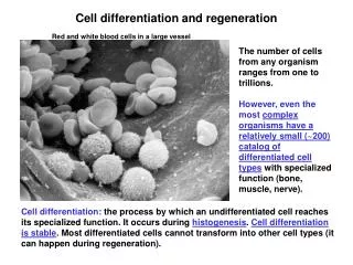
Zygotes - fertilization In living organisms that reproduce sexually, development starts from a single cell, the zygote (dt: befruchtete Eizelle). Zygotes are usually produced by a fertilization event between two haploid cells — an ovum from a female and a sperm cell from a male—which combine to form the single diploid cell. Human sperm and egg (sex cells) have one complete set of chromosomes from the male or female parent. Sex cells, also called gametes, combine to produce somatic cells. Somatic cells therefore have twice as many chromosomes. In humans, gametes have 23 chromosomes. Human somatic cells have 46 chromosomes. www.wikipedia.org Modeling Cell Fate
some terms from developmental biology somatic cells = cells forming the body of an organism germ cells (dt. Keimzelle, Ovolum) are part of the germline. germline (dt. Keimbahn)= line of germ cells that have genetic material that may be passed to a child/embryo. Germline cells are immortal. Gametocyte = eukaryotic germ cell; includes spermatocytes (male) and oocytes (female) primordial germ cells : predecessors of germ cells. They migrate to the gonadal ridge (precursor of gonads). They may be detected from expression of Stella (gene) gonad (dt. Keimdrüse) www.wikipedia.org Modeling Cell Fate
Germ line development Germline cells are produced by embryonic cleavage. Cleavage: division of cells in the early embryo. The zygotes of many species undergo rapid cell cycles with no significant growth. The different cells derived from cleavage are called blastomeres and form a compact mass called the morula (because it resembles a mulberry/ dt. Maulbeere). Cleavage ends with the formation of the blastula. Cleavage in mammals is slow. Cell division takes 12 – 24 hours and is asynchronous. www.wikipedia.org Modeling Cell Fate
From left to right, the morula-stage mouse embryo (embryonic day 2.5; E2.5) holds a core of pre-ICM (inner cell mass) cells that turn into ICM cells at cavitation/ blastulation (E3–E4). At this stage, embryonic stem cell (ESC) and Trophoblast Stem Cell (TSC) cell lines can be derived. As the blastocyst fully expands, the ICM delaminates giving rise to a primitive ectoderm and a primitive endoderm layer. The blastocyst's outer cells are termed trophectoderm. In mammals, the ICM will ultimately form the "embryo proper", while the trophectoderm will form the placenta and other extra-embryonic tissues.[ At E6 and subsequently, the embryo will start gastrulating. This process involves the formation of a mesoderm layer between ectoderm and endoderm, and the formation of the primordial germ cells (PGCs). The pluripotent cells of the embryo are tracked in green. Boiani & Schöler, Nat Rev Mol Cell Biol 6, 872 (2005) Modeling Cell Fate
3 primary germ cell layers The ectoderm is the outer layer of the early embryo. It emerges first and forms from the outer layer of germ cells. The ectoderm differentiates to form the nervous system (spine, peripheral nerves and brain), tooth enamel and the epidermis. It also forms the lining of mouth, anus, nostrils, sweat glands, hair and nails. The endoderm develops at the inner layer. Its cells differentiate to form the gastrointestinal tract, the respiratory tract, endocrine glands and organs, auditory systems, and the urinary system. The mesoderm is the middle layer. It differentiates to give rise to a number of tissues and structures including bone, cartilage (dt: Knorpel), muscle, connective tissue (including that of the dermis), the middle layer of the skin, blood vascular, reproductive, excretory and urinogenital systems and contributes to some glands. www.wikipedia.org Modeling Cell Fate
Developmental Glossary (I) Inner cell mass (ICM): Cells ofthe blastocyst embryo that appear transiently during developmentand give rise to the three germ layers of the developing embryo. Embryonicstem (ES) cells: Pluripotent cell line derived from the ICM uponexplantation in culture. In vitro, ES cells can differentiate intomany different lineages and cell types. Upon injectioninto blastocysts, ES cells can give rise to all tissues including thegermline. Primordial germ cells (PGCs): In vivo, PGCs give rise to oocytesand sperm. When explanted in vitro, PGCs give rise to embryonic germ (EG) cells. Hochedlinger, Development 136, 509 (2009) Modeling Cell Fate
Adult stem cells Embryonic stem cells only exist in the early embryo. We all possess adult stem cells, from which new specialized cells are formed throughout our life time. Adult cells exist predominantly in bone marrow (dt. Knochenmark), but also in skin, fat tissue, umbilical cord (dt. Nabelschnur), brain, liver, and in pancreas (dt. Bauchspeicheldrüse). Adult cells in cell culture have a much reduced ability of self regeneration and a reduced ability for differentiation compared to embryonic stem cells. For example, neural stem cells can differentiate to all cell types of neural tissue (neorons, glia), but likely not into liver or muscle cells. www.wikipedia.org Modeling Cell Fate
Haematopoiesis Haematopoiesis (from Ancient Greek: αἷμα, "blood"; ποιεῖν "to make") is the formation of blood cellular components. All cellular blood compo-nents are derived from haematopoietic stem cells. In a healthy adult person, approximately 1011–1012 new blood cells are produced daily in order to maintain steady state levels in the peripheral circulation. Development of different blood cells from haematopoietic stem cell to mature cells www.wikipedia.org Modeling Cell Fate
Differentiation (Review) A zygote is a eukaryotic cell formed by a fertilization event between two gametes. Zygotes therefore contain DNA derived from both the mother and the father, and this provides all the genetic information necessary to form a new individual. This property is named „totipotency“ (latin: totus – all, potentia – power/ability). Continuous cell division produces daughter cells that start to specialize on individual functions. This developmental process of cells and tissue from a less specialized to a more specialized state is called differentiation in developmental biology. www.wikipedia.org Modeling Cell Fate
Glossary I Totipotency Ability of a cell to giverise to all cells of an organism, including embryonic and extraembryonictissues. Zygotes are totipotent. Pluripotency Ability of a cellto give rise to all cells of the embryo. Cells of the innercell mass (ICM) and its derivative, embryonic stem(ES) cells, are pluripotent. Multipotency Ability of a cellto give rise to different cell types of a given cell lineage.These cells include most adult stem cells, such as gut stemcells, skin stem cells, hematopoietic stem cells and neuralstem cells. Unipotency Capacity of a cell to sustain only onecell type or cell lineage. Examples are terminally differentiatedcells, certain adult stem cells (testis stem cells) and committedprogenitors (erythroblasts). Hochedlinger, Development 136, 509 (2009) Modeling Cell Fate
Epigenetic programming and reprogramming during the mouse life cycle. Two populations of pluripotent cells can be established ex vivo within the time window in which extensive epigenetic reprogramming takes place. These cells are ESCs and embryonic germ cells (EGCs) that are derived from the inner cell mass of the blastocyst and from the PGCs at E8.5–E13.5, respectively. Major remodeling events (e.g. DNA demethylation and X-chromosome reactivation) are highlighted in the figure by colored arrows. TE, trophoectoderm; PE primitive endoderm. Cantone & Fisher, Nature Struct Mol Biol. 20, 292 (2013) Modeling Cell Fate
What is epigenetics? Epigenetics refers to alternate phenotypic states that are not based in differences in genotype, and are potentially reversible, but are generally stably maintained during cell division. Examples: imprinting, twins, cancer vs. normal cells, differentiation, ... Laird, Hum Mol Gen 14, R65 (2005) Modeling Cell Fate
What is epigenetics? A much more expanded view of epigenetics has recently emerged in which multiple mechanisms interact to collectively establish - alternate states of chromatin structure (open – packed/condensed), - histone modifications, • associated protein (e.g. histone) composition, • transcriptional activity, • activity of microRNAs, and - in mammals, cytosine-5 DNA methylation at CpG dinucleotides. Laird, Hum Mol Gen 14, R65 (2005) Modeling Cell Fate
Basic principles of epigenetics:DNA methylation and histone modfications The human genome contains 23 000 genes that must be expressed in specific cells at precise times. Cells manage gene expression by wrapping DNA around clusters (octamers) of globular histone proteins to form nucleosomes. These nucleosomes of DNA and histones are organized into chromatin, the building block of a chromosome. Rodenhiser, Mann, CMAJ 174, 341 (2006) Bock, Lengauer, Bioinformatics 24, 1 (2008) Modeling Cell Fate
Epigenetic modifications Rodenhiser, Mann, CMAJ 174, 341 (2006) Reversible and site-specific histone modifications occur at multiple sites at the unstructured histone tails through acetylation, methylation and phosphorylation. DNA methylation occurs at 5-position of cytosine residues within CpG pairs in a reaction catalyzed by DNA methyltransferases (DNMTs). Modeling Cell Fate
Cytosine methylation Observation: 3-6 % of all cytosines are methylated in human DNA. This methylation occurs (almost) exclusively when cytosine is followed by a guanine base -> CpG dinucleotide. Cytosine 5-methyl-cytosine Mammalian genomes contain much fewer (only 20-25 %) of the CpG dinucleotide than is expected by the G+C content (we expect 1/16 ≈ 6% for any random dinucleotide). This is typically explained in the following way: As most CpGs serve as targets of DNA methyltransferases, they are usually methylated. Esteller, Nat. Rev. Gen. 8, 286 (2007) www.wikipedia.org Modeling Cell Fate
Cytosine methylation 5-Methylcytosine can easily deaminate to thymine. If this mutation is not repaired, the affected CpG is permanently converted to TpG (or CpA if the transition occurs on the reverse DNA strand). Hence, methylCpGs represent mutational hot spots in the genome. If such mutations occur in the germ line, they become heritable. A constant loss of CpGs over thousands of generations can explain the low frequency of this special dinucleotide in the genomes of human and mouse. 5-methyl-cytosine thymine Esteller, Nat. Rev. Gen. 8, 286 (2007) www.wikipedia.org Modeling Cell Fate
effects in chromatin organization affect gene expression Schematic of the reversible changes in chromatin organization that influence gene expression: genes are expressed (switched on) when the chromatin is open (active), and they are inactivated (switched off) when the chromatin is condensed (silent). White circles = unmethylated cytosines; red circles = methylated cytosines. Rodenhiser, Mann, CMAJ 174, 341 (2006) Modeling Cell Fate
Enzymes that controlDNA methylation and histone modfications These dynamic chromatin states are controlled by reversible epigenetic patterns of DNA methylation and histone modifications. Enzymes involved in this process include - DNA methyltransferases (DNMTs), - histone deacetylases (HDACs), - histone acetylases, - histone methyltransferases and the • methyl-binding domain protein MECP2. For example, repetitive genomic sequences (e.g. human endogenous retroviral sequences = HERVs) are heavily methylated, which means transcriptionally silenced. Rodenhiser, Mann, CMAJ 174, 341 (2006) Feinberg AP & Tycko P (2004) Nature Reviews: 143-153 Modeling Cell Fate
DNA methylation Typically, unmethylated clusters of CpG pairs are located in tissue-specific genes and in essential housekeeping genes. (House-keeping genes are involved in routine maintenance roles and are expressed in most tissues.) These clusters, or CpG islands, are targets for proteins that bind to unmethylated CpGs and initiate gene transcription. In contrast, methylated CpGs are generally associated with silent DNA, can block methylation-sensitive proteins and can be easily mutated. The loss of normal DNA methylation patterns is the best understood epigenetic cause of disease. In animal experiments, the removal of genes that encode DNMTs is lethal; in humans, overexpression of these enzymes has been linked to a variety of cancers. Rodenhiser, Mann, CMAJ 174, 341 (2006) Modeling Cell Fate
Differentiation linked to alterations of chromatin structure (B) Upon differentiation, inactive genomic regions may be sequestered by repressive chromatin enriched for characteristic histone modifications. (A) In pluripotent cells, chromatin is hyperdynamic and globally accessible. ML Suva et al. Science 2013; 339:1567-1570 Modeling Cell Fate
Epigenetic stability • In somatic tissues, CpG islands at housekeeping or developmental promoters • are largely unmethylated, whereas non-regulatory CpGs distributed elsewhere • in the genome are largely methylated. • This DNA methylation landscape is relatively static across all somatic tissues. • Most of methylated CpGs are pre-established and inherited through cell division. • In at least two phases of the life cycle of mammals, epigenetic stability is globally perturbed: • when gametes fuse to form the zygote and • when gamete precursors (primordial germ cells; PGCs) develop and migrate in the embryo. • This in vivo ‘reprogramming’ of the epigenetic landscape signals the reacquisition of totipotency in the zygote and the formation of the next generation through PGCs. Cantone & Fisher, Nature Struct Mol Biol. 20, 292 (2013) Modeling Cell Fate
Waddington: Epigenetic landscape Conrad H. Waddington 1956: "Principles of Embryology“; www.nature.com Konrad Hochedlinger and Kathrin Plath, Development 136, 509-523 (2009) Modeling Cell Fate
Epigenetic changes during in vivo reprogramming Global DNA and histone modifi-cations that lead to transcriptional activation of the embryonic genome between the late zygote (paternal genome only) and the 2-cell stage. Protamines are small, arginine-rich, nuclear proteins that replace histones late in the haploid phase of spermatogenesis and are believed essential for sperm head conden-sation and DNA stabilization. In humans, 10-15% of the sperm's genome is packaged by histones thought to bind genes that are essential for early embryonic development (www.wikipedia.org). Gamete genomes undergo different epigenetic programs after fertilization. The paternal genome is mostly subject to epigenetic remodeling at the zygote stage. The maternal genome gradually loses repressive modifications during the subsequent cleavage divisions. Cantone & Fisher, Nature Struct Mol Biol. 20, 292 (2013) Modeling Cell Fate
Epigenetic changes during germline development Global epigenetic changes during germline development from PGC specification (E6.5) to the mitotic/meiotic arrest at E13.5. Two major reprogramming phases can be distinguished during PGC migration toward the genital ridges (E7.5–E10.5) and upon their arrival into the gonads (E10.5–E12.5). Cantone & Fisher, Nature Struct Mol Biol. 20, 292 (2013) Modeling Cell Fate
Hematopoiesis: development of blood cells Orkin & Zon, Cell (2008) 132: 631–644. Modeling Cell Fate
Homework Nature Biotech 33, 269 (2015) The first wave of primitive hematopoiesis originates from Flk1+ mesodermSingle Flk1+ cells were flow sorted at E7.0 (primitive streak, PS), E7.5 (neural plate, NP) and E7.75 (head fold, HF) stages. We subdivided E8.25 cells into putative blood and endothelial populations by isolating GFP+ cells (four somite, 4SG) and Flk1+GFP−cells (4SFG−), respectively Modeling Cell Fate
Summary Epigenetic remodelling is responsible for cellular differentiation. Altering chromatin structure will affect accessibility of genes and, hence, alter the transcriptional program in cells. Open question: - which genes/proteins are the drivers/master regulators? - Does epigenetics regulate transcription, or does transcription regulate epigenetics, or are both closely interlinked? - How can one study such combined epigenetic + gene-regulatory networks by computational modeling? www.wikipedia.org Modeling Cell Fate