Download
1 / 17

E N D
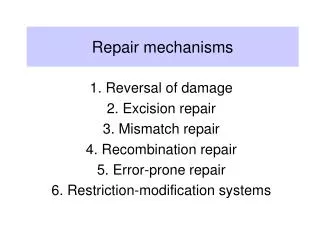
Biological repair mechanisms • There are many potential threats to the fidelity of DNA replication. Not only is there an inherent error rate for the replication of DNA, but there are also spontaneous lesions that can provoke additional errors. Moreover, mutagens in the environment can damage DNA and greatly increase the mutation rate. • Living cells have evolved a series of enzymatic systems that repair DNA damage in a variety of ways. Failure of these systems can lead to a higher mutation rate. A number of human diseases including certain types of cancer can be attributed to defects in DNA repair, as we shall see later. Let's first examine some of the characterized repair pathways and then consider how the cell integrates these systems into an overall strategy for repair. • We can divide repair pathways into several categories.
Prevention of errors before they happen • Some enzymatic systems neutralize potentially damaging compounds before they even react with DNA. One example of such a system is the detoxification of superoxide radicals produced during oxidative damage to DNA: the enzyme superoxide dismutase catalyzes the conversion of the superoxide radicals into hydrogen peroxide, and the enzyme catalase, in turn, converts the hydrogen peroxide into water. Another error-prevention pathway depends on the protein product of the mutT gene: this enzyme prevents the incorporation of 8-oxodG, which arises by oxidation of dGTP, into DNA by hydrolyzing the triphosphate of 8-oxodG back to the monophosphate DNA damage products formed after attack by oxygen radicals. dR = deoxyribose
Direct reversal of damage • The most straightforward way to repair a lesion, once it occurs, is to reverse it directly, thereby regenerating the normal base. Reversal is not always possible, because some types of damage are essentially irreversible. In a few cases, however, lesions can be repaired in this way. One case is a mutagenic photodimer caused by UV light. The cyclobutane pyrimidine photodimer can be repaired by a photolyase that has been found in bacteria and lower eukaryotes but not in humans. The enzyme binds to the photodimer and splits it, in the presence of certain wavelengths of visible light, to generate the original bases. This enzyme cannot operate in the dark, so other repair pathways are required to remove UV damage. A photolyase that reverses the 6-4 photoproducts has been detected in plants and Drosophila. Repair of a UV-induced pyrimidine photodimer by a photoreactivating enzyme, or photolyase. The enzyme recognizes the photodimer (here, a thymine dimer) and binds to it. When light is present, the photolyase uses its energy to split the dimer into the original monomers.
Removal of alkyl groups • Alkyltransferases also are enzymes taking part in the direct reversal of lesions. They remove certain alkyl groups that have been added to the O-6 positions of guanine by such agents as NG and EMS. The methyltransferase from E. coli has been well studied. This enzyme transfers the methyl group from O-6-methylguanine to a cysteine residue on the protein. When this happens, the enzyme is inactivated, so this repair system can be saturated if the level of alkylation is high enough. Alkylation-induced specific mispairing. The alkylation (in this case, EMS-generated ethylation) of the O-6 position of guanine andthe O-4 position of thymine can lead to direct mispairing with thymine and guanine, respectively, as shown here. In bacteria, where mutations have been analyzed in great detail, the principal mutations detected are GC → AT transitions, indicating that the O-6 alkylation of guanine is most relevant to mutagenesis.
Excision-repair pathways Also termed nucleotide excision repair, this system includes the breaking of a phosphodiester bond on either side of the lesion, on the same strand, resulting in the excision of an oligonucleotide. This excision leaves a gap that is filled by repair synthesis, and a ligase seals the breaks. In prokaryotes, 12 or 13 nucleotides are removed; whereas, in eukaryotes, from 27 to 29 nucleotides are eliminated.
The excinuclease • In E. coli, the products of the uvrA, B, and C genes constitute the excinuclease. The UvrA protein, which recognizes the damaged DNA, forms a complex with UvrB and leads the UvrB subunit to the damage site before dissociating. The UvrC protein then binds to UvrB. Each of these subunits makes an incision. The short DNA 12-mer is unwound and released by another protein, helicase II. The human excinuclease is considerably more complex than its bacterial counterpart and includes at least 17 proteins. However, the basic steps are the same as those in E. coli Schematic representation of events following incision by UvrABC exinuclease in E. coli.
Excision repair defects in humans • Several human genetic diseases are known to be due to repair defects. • Xeroderma pigmentosum (XP) results from a defect in any of the genes (complementation groups) effecting nucleotide excision repair. People suffering from this disorder are extremely prone to UVinduced skin cancers as a result of exposure to sunlight and have frequent neurological abnormalities. Nucleotide excision repair is coupled to transcription. This model for coupled repair in mammalian cells shows RNA polymerase (pink) pausing when encountering a lesion. It undergoes a conformational change, allowing the DNA strands at the lesion site to reanneal. Protein factors aid in coupling by bringing TFIIH and other factors to the site to carry out the incision, excision, and repair reactions. Then transcription can continue normally.
Specific excision pathways • Certain lesions are too subtle to cause a distortion large enough to be recognized by the uvrABC-encoded general excision-repair system and its counterparts in higher cells. Thus, additional excision pathways are necessary. • DNA glycosylase repair pathway (base-excision repair).DNA glycosylases do not cleave phosphodiester bonds, but instead cleave N-glycosidic (base–sugar) bonds, liberating the altered base and generating an apurinic or an apyrimidinic site, both called AP sites, because they are biochemically equivalent. The resulting Ap site is then repaired by an AP endonuclease repair pathway
Mismatch repair • Some repair pathways are capable of recognizing errors even after DNA replication has already occurred. One such system, termed the mismatch repair system, can detect mismatches that occur in DNA replication. Suppose you were to design an enzyme system that could repair replication errors. What would this system have to be able to do? At least three things: 1. Recognize mismatched base pairs. 2. Determine which base in the mismatch is the incorrect one. 3. Excise the incorrect base and carry out repair synthesis. • The second point is the crucial property of such a system. Unless it is capable of discriminating between the correct and the incorrect bases, the mismatch repair system could not determine which base to excise. If, for example, a G–T mismatch occurs as a replication error, how can the system determine whether G or T is incorrect? Both are normal bases in DNA. But replication errors produce mismatches on the newly synthesized strand, so it is the base on this strand that must be recognized and excised.
DNA methylation • To distinguish the old, template strand from the newly synthesized strand, the mismatch repair system in bacteria takes advantage of the normal delay in the postreplication methylation of the sequence The methylating enzyme is adenine methylase, which creates 6-methyladenine on each strand. However, it takes the adenine methylase several minutes to recognize and modify the newly synthesized GATC stretches. During that interval, the mismatch repair system can operate because it can now distinguish the old strand from the new one by the methylation pattern. Methylating the 6-position of adenine does not affect base pairing, and it provides a convenient tag that can be detected by other enzyme systems.
Mismatch repair Model for mismatch repair in E. coli. Because DNA is methylated by enzymatic reactions that recognize the A in a GATC sequence, the newly synthesized strand will not be methylated directly after DNA replication. The hemimethylated DNA duplex serves as a recognition point for the mismatch repair system in discerning the old from the new strand. Here a G–T mismatch is shown. The mismatch repair system can recognize and bind to this mismatch, determine the correct (old) strand because it is the methylated strand of a hemimethylated duplex, and then excise the mismatched base from the new strand. Repair synthesis restores the normal base pair.
The complex MutS-MutH • Steps in E. coli mismatch repair. • (1) MutS binds to mispair. • (2) MutH and MutL are recruited to form a complex. MutH cuts the newly synthesized (unmethylated) strand, and exonuclease degradation goes past the point of the mismatch, leaving a patch. • (3) Single-strand-binding protein (Ssb) protects the single-stranded region across from the missing patch. • (4) Repair synthesis and ligation fill in the gap.
Mismatch repair defects in humans Hereditary nonpolyposis colorectal cancer (HNPCC) is one of the most common inherited predispositions to cancer, affecting as many as 1 in 500 people in the Western world. Most HNPCC results from a defect in genes that encode the human counterparts (and homologs) of the bacterial MutS and MutL proteins. The inheritance of HNPCC is autosomal dominant. Cells with one functional copy of the mismatch repair genes have normal mismatch repair activity, but tumor cell lines arise from cells that have lost the one functional copy and are thus mismatch repair deficient. These cells display high mutation rates that eventually result in tumor growth and proliferation. Mismatch repair in humans. (1) Mispairs and misaligned bases arise in the course of replication. (2) The G–T-binding protein (GTBP) and the human MutS homolog (hMSH2) recognize the incorrect matches. (3) Two additional proteins, hPMS2 and hMLH1, are recruited and form a larger repair complex. (4) The mismatch is repaired after removal, DNA synthesis, and ligation.
SOS repair DNA damage often results ina replication block, because DNA synthesis will not proceed past a base that cannot specify its complementary partner by hydrogen bonding. In bacterial cells, such replication blocks can be bypassed by inserting nonspecific bases. The process requires the activation of a special system, the SOS system. The name SOS comes from the idea that this system is induced as an emergency response to prevent cell death in the presence of significant DNA damage. SOS induction is a last resort, allowing the cell to trade death for a certain level of mutagenesis. The recA gene, takes part in postreplication repair. Here the DNA replication system stalls at a UV photodimer and then restarts past the block, leaving a single-stranded gap. This process leads to few errors. SOS bypass, in contrast, is highly mutagenic. Here the replication system continues past the lesion, accepting noncomplementary nucleotides for new strand synthesis
Schemes for postreplication repair (a) In recombinational repair, replication jumps across a blocking lesion, leaving a gap in the new strand. A recA-directed protein then fills the gap, using a piece from the opposite parental strand (because of DNA complemen-tarity, this filler will supply the correct bases for the gap). Finally, the RecA protein repairs the gap in the parental strand. (b) In SOS bypass, when replication reaches a blocking lesion, the SOS system inserts the necessary number of bases (often incorrect ones) directly across from the lesion and replication continues without a gap. Note that with either pathway the original blocking lesion is still there and must be repaired by some other repair pathway.
Summary of repair mechanisms • We can now assess the overall repair system strategy used by the cell. It would be convenient if enzymes could be used to directly reverse each specific lesion. However, sometimes that is not chemically possible, and not every possible type of DNA damage can be anticipated. • Therefore, a general excision repair system is used to remove any type of damaged base that causes a recognizable distortion in the double helix. • When lesions are too subtle to cause such a distortion, specific excision systems, glycosylases, or removal systems are designed. • To eliminate replication errors, a postreplication mismatch repair system operates; finally, postreplication recombinational systems eliminate gaps across from blocking lesions that have escaped the other repair systems. • The SOS system is the last resort for a cell to survive a potentially lethald DNA damage
DNA repair and mutation rates • The repair processes are so efficient that the observed base substitution rate is as low as 10−10 to 10−9 per base pair per cell per generation in E. coli. However, mutant strains with increased spontaneous mutation rates have been detected. Such strains are termed mutators. In many cases, the mutator phenotype is due to a defective repair system. In humans, these repair defects often lead to serious diseases. • In E. coli, the mutator loci mutH, mutL, mutU, and mutS affect components of the postreplication mismatch repair system, as does the dam locus, which specifies the enzyme deoxyadenosine methylase. Strains that are Dam− cannot methylate adenines at GATC sequences, and so the mismatch repair system can no longer discriminate between the template and the newly synthesized strands. This failure to discriminate leads to a higher spontaneous mutation rate. • Mutations in the mutY locus result in GC → TA transversions, because many G–A mispairs and all 8-oxodG–A mispairs are unrepaired. The mutM gene encodes a glycosylase that removes 8-oxodG. Strains lacking mutM are mutators for the GC → TA transversion. Strains that are MutT− have elevated rates of the AT → CG transversion, because they lack an activity that prevents the incorporation of 8-oxodG across from adenine. • Strains that are Ung− are missing the enzyme uracil DNA glycosylase. These mutants cannot excise the uracil resulting from cytosine deaminations and, as a result, have elevated levels of C → T transitions. The mutD locus is responsible for a very high rate of mutagenesis (at least three orders of magnitude higher than normal). Mutations at this locus affect the proofreading functions of DNA polymerase III.