Download
1 / 24
320 likes | 482 Vues
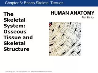
The skeletal system provides support to the body, protects vital organs, and enables movement through attached skeletal muscles. It also serves as a reservoir for minerals and fats and plays a crucial role in blood cell formation. This system consists of bones, joints, cartilages, and ligaments, categorized into the axial and appendicular skeleton. The adult human skeleton comprises 206 bones, classified into four types based on shape: long, short, flat, and irregular bones. Each bone type has unique anatomical features, contributing to the system's complexity and functionality.

E N D
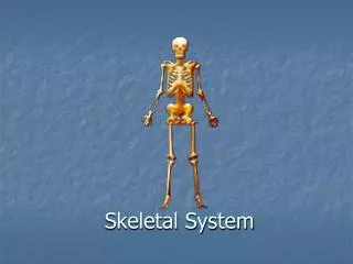
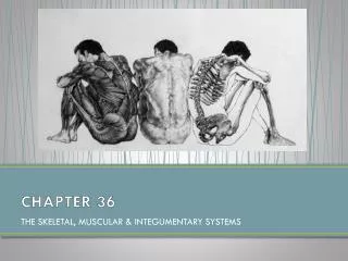
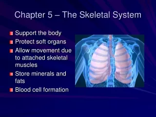
Chapter 5 – The Skeletal System • Support the body • Protect soft organs • Allow movement due to attached skeletal muscles • Store minerals and fats • Blood cell formation
The Skeletal System • Parts of the skeletal system • Bones (skeleton) • Joints • Cartilages • Ligaments • Two subdivisions of the skeleton • Axial skeleton • Appendicular skeleton
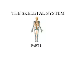
Bones of the Human Body • The adult skeleton has 206 bones • Two basic types of bone tissue • Compact bone • Spongy bone
Classification of Bones • Long bones • Typically longer than they are wide • Have a shaft with heads at both ends • Contain mostly compact bone • Example: • Femur • Humerus Shaft
Classification of Bones • Short bones • Generally cube-shape • Contain mostly spongy bone • Example: • Carpals • Tarsals
Classification of Bones • Flat bones • Thin, flattened, and usually curved • Two thin layers of compact bone surround a layer of spongy bone • Example: • Skull • Ribs • Sternum
Classification of Bones • Irregular bones • Irregular shape • Do not fit into other bone classification categories • Example: • Vertebrae • Hip bones
Anatomy of a Long Bone • Diaphysis • Shaft • Composed of compact bone • Epiphysis • Ends of the bone • Composed mostly of spongy bone
Anatomy of a Long Bone • Periosteum • Outside covering of the diaphysis (shaft) • Fibrous connective tissue membrane
Anatomy of a Long Bone • Articular cartilage • Covers the external surface of the epiphyses • Made of hyaline cartilage • Decreases friction at joint surfaces
Anatomy of a Long Bone • Epiphyseal plate • Flat plate of hyaline cartilage seen in young, growing bone (a.k.a. = growth plate) • Epiphyseal line • Remnant of the epiphyseal plate • Seen in adult bones
Anatomy of a Long Bone • Medullary cavity • Cavity inside of the shaft • Contains yellow marrow (mostly fat) in adults • Contains red marrow (for blood cell formation) in infants
Bone Markings • Surface features of bones • Sites of attachments for muscles, tendons, and ligaments • Passages for nerves and blood vessels • Categories of bone markings • Projections or processes — grow out from the bone surface • Depressions or cavities — indentations
Bone Surface Markings • projections where muscles, tendons, ligaments attach • tuberosity – large rounded projection • spinous process (or spine) – sharp, slender, pointed projection • trochanter – very large, blunt irregular shaped projection • crest – narrow ridge of bone
Bone Surface Markings • Projections that help to form joints • Head – bony expansion at the end of a long neck • Facet – smooth, almost flat surface • Condyle – rounded projection • Ramus – armlike arm of bone
Bone Surface Markings • Depressions and openings for blood vessels and nerves to pass through • Foramen – round or oval opening in a bone • Meatus – canal-like • Fossa – shallow depression mostly to form a joint
Microscopic Anatomy of Bone • Osteon (Haversian system) • A unit of bone containing central canal and matrix rings • Central (Haversian) canal • Opening in the center of an osteon • Carries blood vessels and nerves • Perforating (Volkman’s) canal • Canal perpendicular to the central canal • Carries blood vessels and nerves
Microscopic Anatomy of Bone Figure 5.3a
Microscopic Anatomy of Bone • Lacunae • Cavities containing bone cells (osteocytes) • Arranged in concentric rings • Lamellae • Rings around the central canal • Sites of lacunae
Microscopic Anatomy of Bone Figure 5.3b–c
Microscopic Anatomy of Bone • Canaliculi • Tiny canals • Radiate from the central canal to lacunae • Form a transport system connecting all bone cells to a nutrient supply
Microscopic Anatomy of Bone Figure 5.3b
Types of Bone Cells • Osteocytes—mature bone cells • Osteoblasts—bone-forming cells • Osteoclasts—bone-destroying cells • Break down bone matrix for remodeling and release of calcium in response to parathyroid hormone • Bone remodeling is performed by both osteoblasts and osteoclasts