Download
1 / 19
260 likes | 757 Vues
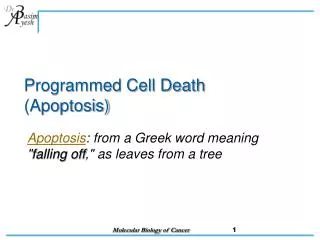
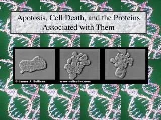
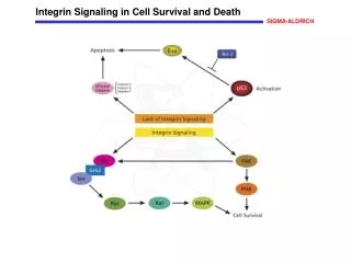
Signaling to PROGRAM cell death (Apoptosis). Apoptosis is a cell mechanism used to eliminate cells that are unnecessary to or that contain mutations that are dangerous to the body . Loss of the ability to undergo apoptosis leads to cancer. . 1. Phenotypes of apoptosis:

E N D
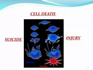
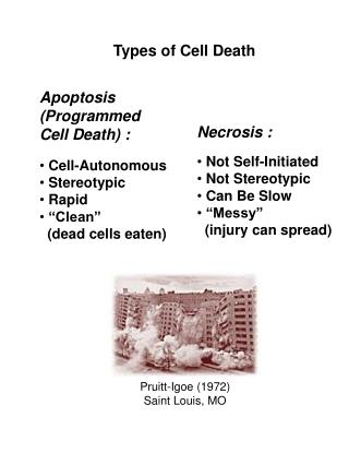
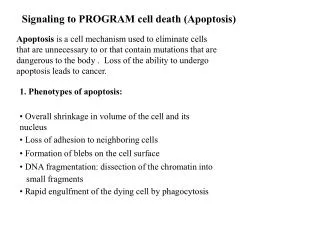
Signaling to PROGRAM cell death (Apoptosis) Apoptosis is a cell mechanism used to eliminate cells that are unnecessary to or that contain mutations that are dangerous to the body . Loss of the ability to undergo apoptosis leads to cancer. 1. Phenotypes of apoptosis: • Overall shrinkage in volume of the cell and its nucleus • Loss of adhesion to neighboring cells • Formation of blebs on the cell surface • DNA fragmentation: dissection of the chromatin into small fragments • Rapid engulfment of the dying cell by phagocytosis
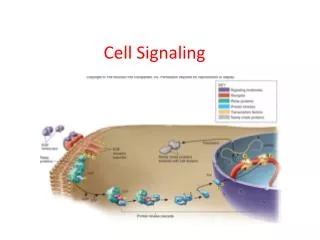
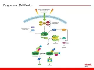
Apoptosis (Program cell death) • Factors that induce apoptosis: Internal stimuli: abnormalities in DNA External stimuli: removal of growth factors,addition of cytokines (Tumor Necrosis Factor or TNF) Signal transduction pathways leading to apoptosis: There are two major pathways: intrinsic pathway (mitochondria-dependent) and extrinsic pathway (mitochondria-independent). •
Death receptor pathway • The death receptor pathway is activated by external cytokines and is mitochondria-independent • The ligands of the death receptors are members of the tumor necrosis factor (TNF) family of proteins, including TNFa, Fas ligand (FasL), TRAIL/Apo2L, Apo3L FasL TRAIL TNFa Apo3L
Activation of the death receptors • Binding of ligand to the death receptors results in homo trimerization of the receptors • The death receptors contain a death domain in the cytoplasmic region that is required for apoptosis signaling
Activation of the death receptors • Trimerization of the receptor death domains allows binding and activation of FADD (Fas-associated death domain protein) and formation of death-inducing signaling complex (DISC) DISC recruits and activates Procaspase 8 and 10
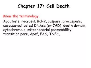
Activation of procaspases • Caspases are a family of cysteine aspartyl-specific proteases that are activated at an early stage of apoptosis and are responsible for triggering most, if not all, of the changes observed during apoptosis. • In the absence of stimulation, caspases exist in the cell as an inactive precursor. Activation of caspases by DISC results in enzymatic removal of portions of the caspase precursors and release of smaller fragments that are active. inactive active The active caspase then diffuses into the cytoplasm and cleaves target proteins. •
Two major classes of caspases • Initiator caspases: initiate the onset of apoptosis by activating the executioner caspases Caspase 8, caspase 9, caspase 10, etc. • Executioner caspases: destroy actual targets in the cell to execute apoptosis Caspase 3, caspase 6, caspase 7
Some cellular targets of caspases FAK (focal adhesion kinase): inactivation of FAK disrupt cell adhesion, leading to detachment of the apoptotic cell from its neighbors. Lamins: important component of the nuclear envelope. Cleavage of lamins leads to disassembly of the nuclear lamina. Proteins required for cell structure: actin, intermediate filaments, etc. Cleavage of these proteins lead to changes in cell shape and the surface blebbing. Endonuclease CAD:responsible for chromosome fragmentation. CAD cuts DNA into small fragments. CAD normally binds to an inhibitor protein. Caspases cleaves the inhibitor protein and activates CAD. Enzymes involved in DNA repair: no need to repair DNA in cells destined to death.
procaspase 3 caspase 3 Summary of death receptor pathway ligands Death receptors FADD/DISC Caspase 8, 10 Caspase 3, 6, 7 Lamin, CAD,actin, Adhesion molecules, etc
Apoptosis (Program cell death) • Factors that induce apoptosis: Internal stimuli: abnormalities in DNA External stimuli: removal of growth factors,addition of cytokines (Tumor Necrosis Factor or TNF) Signal transduction pathways leading to apoptosis: There are two major pathways: intrinsic pathway (mitochondria-dependent) and extrinsic pathway (mitochondria-independent). •
Mitochondria The outer membrane and intermembrane space are involved in apoptosis signaling
The bcl-2 family of proteins 6 proteins 18 proteins BH: bcl-2 homology
The bcl-2 family proteins homo- and hetero-dimerize • The Bcl-2 family members can form homo- or hetero-dimers through the BH3 domains. • The pro-apoptotic Bax homo-dimers promotes apoptosis. • Bcl-2 forms hetero-dimers with Bax, leading to the inhibition of the apoptotic activity of Bax. Thus, the relative levels of pro-survival and pro-apoptosis Bcl-2 family proteins determine cell survival or apoptosis. How do these proteins regulate apoptosis?
cytoplasm intermembrane space Bcl-2 family proteins control cytochrome c translocation • Bax forms homo-dimers in the presence of apoptotic signals. This homodimer presumably promotes the opening of a channel that controls the translocation of cytochrome c from the intermembrane space to cytoplasm • Bcl-2 interferes with Bax function by forming a hetero dimer with Bax. This leads to the closing of the channel and inhibition of cytochrome c translocation
Cytochrome c translocation initiates caspase cascade Apaf-1 • In the cytosol, cytochrome c binds to Apaf-1 to form apoptosome • The apoptosome recruits procaspase 9 to generate the active caspase 9 Cyt. c • Caspase 9 then activates the executioner caspase 3, 6, 7 Procaspase 9 Caspase 9 Apoptosome Procaspase 3 Caspase 3
Extrinsic and intrinsic pathways converge at executioner caspases ligands Intrinsic apoptosis signals Pro-apoptotic Bcl-2 proteins Death receptors Cytochrome c translocation FADD/DISC Apoptosome (Apaf-1/cyt.c) Caspase 8, 10 Caspase 9 Caspase 3, 6, 7 Lamin, CAD,actin, Adhesion molecules, etc
Apoptosis lab: Induction of apoptosis of Jurkat human T-cells by staurosporine. Caspase assay--measure caspase activation Activation of caspase 3 results in the cleavage of a synthetic caspase 3 substrate called DEVD-pNA (aspartate-glutamate-valine-aspartate p-nitroanalide). Cleavage of DEVD-pNA releases pNA, which can then be detected by a change it its absorbance at 405 nm. TUNEL assay--measure DNA fragmentation
3’ 5’ TdT FITC-dUTP 3’ 5’ Methods to measure apoptosis TUNEL assay: measure DNA fragmentation TdT (terminal deoxynucleotidyl transferase): adds dNTPs to the 3’ end of DNA molecules in the absence of a template Bright field FITC