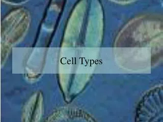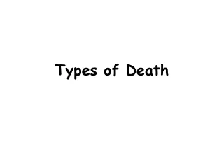
Types of Cell Death
210 likes | 544 Vues
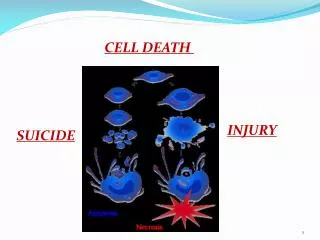
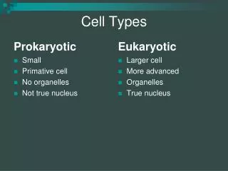
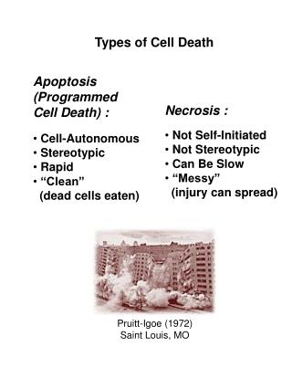
Apoptosis (Programmed Cell Death) :. Necrosis :. Not Self-Initiated Not Stereotypic Can Be Slow “Messy” (injury can spread). Cell-Autonomous Stereotypic Rapid “Clean” (dead cells eaten). Pruitt-Igoe (1972) Saint Louis, MO. Types of Cell Death. Questions :

Types of Cell Death
E N D
Presentation Transcript
Apoptosis (Programmed Cell Death) : Necrosis : • Not Self-Initiated • Not Stereotypic • Can Be Slow • “Messy” • (injury can spread) • Cell-Autonomous • Stereotypic • Rapid • “Clean” • (dead cells eaten) Pruitt-Igoe (1972) Saint Louis, MO Types of Cell Death
Questions : • Is Apoptosis a “real” biological • phenomenon (controlled by a genetic • program)? • How are cells killed? • What turns apoptosis on and off? • Can this program be altered for • good or evil? • i.e. cancer…too little apoptosis • or neurodegenerative diseases • …too much apoptosis
Lecture to Cover: • Classic experiments describing • the phenomenology of cell death. • Identification of NGF and other • protein trophic factors. • Cloning of genes comprising the • death mechanism. • Molecular model for apoptosis.
Experimental removal of neuronal target cells, results in excessive loss of projecting neurons. Deprived Control (after Hamburger, 1958, 1977)
Massive loss of neurons inembryos occurs during normal development. Lateral Motor Column (40% Loss) Ciliary Ganglion (54% Loss) Trochlear Nucleus (57% Loss) (after Hamburger, 1975; Landmesser and Pilar, 1974; Cowan and Wenger, 1967)
Increasing Developmental Time Loss of neurons timed with innervation of target muscles. Is there a relationship? Target Innervation Cell Loss
Increasing Developmental Time Extra target cells (more neurons) Fewer target cells (fewer neurons) Number of target cells determines the number of innervation neurons that survive.
Mouse sarcoma transplanted next to developing chick nerve cord causes extra sprouting of neurons. (Diffusible factor suggested) (Levi-Montalcini and Hamburger, 1951)
Development of a quantitative functional assay for nerve growth factor (NGF) activity, using explanted cultures of sensory ganglia. (-NGF) (Levi-Montalcini, Hamburger and Cohen, 1954) (+NGF) Rita Levi-Montalcini Viktor Hamburger Stanley Cohen
NGF is the founding member of a large gene family of neurotrophic proteins, distantly related to insulin. Neurotrophin Gene Family
NGF/Neurotrophins Signal Through Trk (tyrosine kinase) Receptors. NGF/NT Trk Receptor Kinases? Apoptosis pathway? Transcription Factor? Gene Activation/ Repression
Bodywall Muscle Hypoderm Neurons Cuticular Cells Pharynx developmental time Intestine Vulva Gonad Germ Cells Muscle Neuronal Cell Death Lineages (Brenner, 1973; Sulston and Horvitz, 1977;White, Horvitz, Sulston, 1982; Sulston, Schierenberg, White, Thomson, 1983 ) Sydney Brenner John Sulston H. Robert Horvitz C. elegans is a great model organism for molecular genetic studies of Cell Death.
Programmed Cell Death of single identified cells can be observed in live worms. P11aap (Sulton and Horvitz, 1977)
WT (pro-survival genes + pro-apoptosis genes) 2 Classes of C. elegans Cell Death Mutants (normal number of cells) Mutant class I (pro-survival genes + pro-apoptosis genes) (fewer cells) Mutant class II (pro-survival genes + pro-apoptosis genes) (extra cells)
ced-4 ced-9 ced-3 Genetic analysis of cell death genes in C. elegans defines a genetic pathway. Cell Death (pro-survival) gene (pro-apoptosis) genes
Bcl-2 Chromosome 18 Ig Heavy Chain Chromosome 14 Bcl-2 Chromosome 18 Ig Heavy Chain Chromosome 14 Bcl-2 t(14;18) Chromosomal Translocation Stanley Korsmeyer t(14;18) Chromosomal Translocation Causes Human B-cell Leukemia by Overexpression of Bcl-2. (from Korsmeyer, et al.; Croce, et al.; Sklar, et al.; 1985 - 1990)
ced-9 / Bcl-2: • Bcl-2: B-cell Leukemia. • “Pro-survival” protein. • Inhibits release of • cytochrome C from • mitochondria (vertebrates). • Sequesters CED-4 • from cytoplasm (worms). Ce Ce Ce Hu Hu Hu ced-4 / Apaf: • Apaf:Apoptosis Activity Factor. • “Adaptor” or “scaffold” protein. • Aggregates inactive procaspase, • causing auto-activation by proximity. • Requires cytochrome C, and ATP • for multimerization (vertebrates). ced-3 / Caspase: • Caspase:Active-site • Cysteine, Aspartate • Protease. • “Terminator” protein. • Protease activity • when activated by • proteolysis. The “Core” Cell Death genes found in C. elegans are evolutionarily conserved as multigene families in vertebrates.
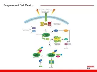
mitochondria Bcl-2 (ced-9) single BH3 domain protein (egl-1) - - - (BH3 domains) Apaf (ced-4) Cytochrome C caspase (ced-3) (procaspase recruitment) - - (Catalysis of the removal of self-inhibitory caspase domain) - - - - Apaf aggregation Recruitment of inactive procaspase activated caspase (cascade) Death Molecular Model for Apoptosis
NGF maybe one of multiple pathways to the “core” death mechanism, mediated by many single-BH3 proteins. PDGF NGF Single BH3 domain protein Single BH3 domain protein Single BH3 domain protein (From Gross, McDonnell, and Korsmeyer, 1999)
BID (single-BH3) Bcl-xL (Bcl-2 like) Diptheria Toxin (pore forming) The C. elegansegl-1 gene resembles vertebrate single BH3 domain molecules that trigger apoptosis. Mitochondria integrate “Pro-survival” and “Pro-death” signals from a family of Bcl-2-like genes. Pro-survival Pro-death Bcl-2 Bax Bik (BH3) (BH3) “Pro-death” Single-BH3 domain proteins complex with Bcl-2 to release cytochrome C from mitochondria. + BH3 BH3 (from Fesik, 2000)
WD-40 Apaf WD-40 CARD (caspase activation and recruitment domain) Cytochrome C Apaf gene +procaspase-9 (x7?) pc-9 Apaf/Cytochrome C Aggregate into a 7-Spoke Apoptosome Complex (Acehan, et al., 2002)