Download
1 / 123
1.24k likes | 1.34k Vues
Discover the causes of cell injury, from genetic derangements to oxygen deprivation, and learn how reversible and irreversible damage affects cells in the body, impacting overall health.

E N D
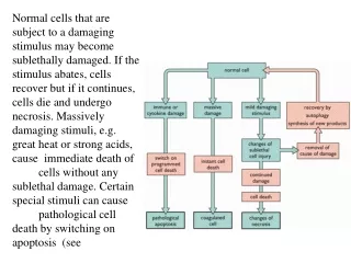
Normal cells that are subject to a damaging stimulus may become sublethally damaged. If the stimulus abates, cells recover but if it continues, cells die and undergo necrosis. Massively damaging stimuli, e.g. great heat or strong acids, cause immediate death of cells without any sublethal damage. Certain special stimuli can cause pathological cell death by switching on apoptosis (see
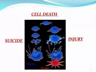
Cell death, • is one of the most crucial events in the evolution of disease of any tissue or organ.
Causes of cell injury • External causes • gross physical violence of an automobile accident to • internal endogenous causes, • a subtle genetic mutation causing lack of a vital enzyme that impairs normal metabolic function.
Physical Agents. • Prick of a thorn • mechanical trauma, • extremes of temperature (burns and deep cold), • sudden changes in atmospheric pressure, • radiation, • electric shock
Chemical Agents and Drugs • Simple chemicals such as glucose or salt in hypertonic concentrations may cause cell injury directly or by deranging electrolyte balance • Even oxygen, in high concentrations, is severely toxic. • Trace amounts of agents known as poisons, such as arsenic, cyanide, or mercuric salts, may destroy sufficient numbers of cells within minutes to hours to cause death.
Chemical Agents and Drugs • environmental and air pollutants, • insecticides, and herbicides; • industrial and occupational hazards, such as carbon monoxide and asbestos; • social stimuli, such as alcohol and narcotic drugs; • variety of therapeutic drugs.
Infectious Agents. • viruses • tapeworms. rickettsiae, • bacteria, • fungi,
Immunologic Reactions. • immune system serves an essential function in defense against infectious pathogens, • immune reactions may, in fact, cause cell injury. • The anaphylactic reaction to a foreign protein or a drug is a prime example, • reactions to endogenous self-antigens are responsible for a number of autoimmune disease
Oxygen Deprivation. • Hypoxia is a deficiency of oxygen, which causes cell injury by reducing aerobic oxidative respiration. • Hypoxia is an extremely important and common cause of cell injury and cell death.
hypoxia is inadequate oxygenation • cardiorespiratory failure. • Loss of the oxygen-carrying capacity of the blood, as in anemia or • carbon monoxide poisoning (producing a stable carbon monoxyhemoglobin that blocks oxygen carriage), • Depending on the severity of the hypoxic state, cells may adapt, undergo injury, or die. • if the femoral artery is narrowed, the skeletal muscle cells of the leg may shrink in size (atrophy). • a balance between metabolic needs and the available oxygen supply may be achieved. More severe hypoxia induces injury and cell death.
ischemia • loss of blood supply from impeded arterial flow or reduced venous drainage in a tissue. • Ischemia compromises the supply not only of oxygen, but also of metabolic substrates, including glucose (normally provided by flowing blood). • ischemic tissues are injured more rapidly and severely than are hypoxic tissues.
Genetic Derangements. • genetic injury may result in a defect as severe as the congenital malformations associated with Down syndrome, caused by a chromosomal abnormality, • subtle as the decreased life of red blood cells caused by a single amino acid substitution in hemoglobin S in sickle cell anemia. • Variations in the genetic makeup can also influence the susceptibility of cells to injury by chemicals and other environmental insults.
Nutritional Imbalances. • Protein-calorie deficiencies • Deficiencies of specific vitamins • Nutritional problems can be self-imposed • as in anorexia nervosa or • self-induced starvation. • nutritional excesses have also become important causes of cell injury.
Nutritional Imbalances. • Excesses of lipids predispose to atherosclerosis, • obesity is a manifestation of the overloading of some cells in the body with fats. • In addition to the problems of under nutrition and over nutrition, the composition of the diet makes a significant contribution to a number of diseases. • Metabolic diseases such as diabetes also cause severe cell injury.
Reversible injury • generalized swelling of the cell and its organelles; • blebbing of the plasma membrane; • detachment of ribosomes from the endoplasmic reticulum; • and clumping of nuclear chromatin. • Laminated structures (myelin figures) derived from damaged membranes of organelles and the plasma membrane appear
irreversible injury • increasing swelling of the cell; • disruption of cellular membranes; • swelling and disruption of lysosomes; • presence of large amorphous densities in swollen mitochondria; • and profound nuclear changes. • The latter include nuclear codensation (pyknosis), • followed by fragmentation (karyorrhexis) • and dissolution of the nucleus (karyolysis). • Laminated structures (myelin figures) become more pronounced in irreversibly damaged cells.
Proverb of this week Necessity is the mother of invention
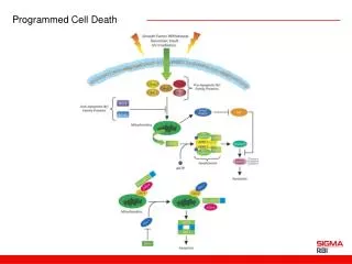
There are two principal patterns of cell death, necrosis and apoptosis.
cell death the morphological pattern of which is called necrosis occurs after such abnormal stresses as ischemia and chemical injury, and it is always pathological.
Apoptosis occurs when a cell dies through activation of an internally controlled suicide program.
Physiological • It is designed to eliminate unwanted cells during embryogenesis • and in various physiologic processes, such as involution of hormone-responsive tissues upon withdrawal of the hormone.
Pathological apoptosis • Immunological injuries
9 The sequential ultrastructural changes seen in necrosis (left) and apoptosis (right). In apoptosis, the initial changes consist of nuclear chromatin condensation and fragmentation, followed by cytoplasmic budding and phagocytosis of the extruded apoptotic bodies.
Mechanism of injury • The cellular response to injurious stimuli depends on • the type of injury, • its duration,and • its severity • depend on • the type, • state, • and adaptability of the injured cell. • The cell's nutritional and hormonal status • and its metabolic needs.
Mechanism of injury • How vulnerable is a cell, for example, to loss of blood supply and hypoxia? • The striated muscle cell is less vulnerable to deprivation of its blood supply; not so the striated muscle of the heart. • Exposure of two individuals to identical concentrations of a toxin, such as carbon tetrachloride, may produce no effect in one and cell death in the other. • This may be due to genetic variations affecting the amount and activity of hepatic enzymes that convert carbon tetrachloride to toxic byproducts
targets of injury • aerobic respiration involving mitochondrial oxidative phosphorylation and production of ATP; • the integrity of cell membranes, on which the ionic and osmotic homeostasis of the cell and its organelles depends; • protein synthesis; • the integrity of the genetic apparatus of the cell
Mechanism of injury • DEPLETION OF ATP • MITOCHONDRIAL DAMAGE • INFLUX OF CALCIUM AND LOSS OF CALCIUM HOMEOSTASIS • ACCUMULATION OF OXYGEN-DERIVED FREE RADICALS (OXIDATIVE STRESS) • DEFECTS IN MEMBRANE PERMEABILITY
DEPLETION OF ATP Functional and morphologic consequences of decreased intracellular ATP during cell injury.
INFLUX OF INTRACELLULAR CALCIUM AND LOSS OF CALCIUM HOMEOSTASIS Sources and consequences of increased cytosolic calcium in cell injury. ATP, adenosine triphosphate.
ACCUMULATION OF OXYGEN-DERIVED FREE RADICALS (OXIDATIVE STRESS)
Reversible vs. irreversible cell injury • there are clearly many ways to injure a cell, • the "point of no return," at which irreversible damage has occurred, is still largely undetermined; • thus, we have no precise cut-off point • dissolution of the injured cell is characteristic of necrosis, one of the recognized patterns of cell death. • There is also widespread leakage of potentially destructive cellular enzymes into the extracellular space, with damage to adjacent tissues
Reversible vs. irreversible cell injury • leakage of intracellular proteins across the degraded cell membrane into the peripheral circulation provides a means of detecting tissue-specific cellular injury and death using blood serum samples. • Cardiac muscle, for example, contains a specific isoform of the enzyme creatine kinase and of the contractile protein troponin; • liver (and specifically bile duct epithelium) contains a temperature-resistant isoenzyme of the enzyme alkaline phosphatase; • and hepatocytes contain transaminases. • Irreversible injury and cell death in these tissues are consequently reflected in increased levels of such proteins in the blood.