Download
1 / 8
100 likes | 734 Vues
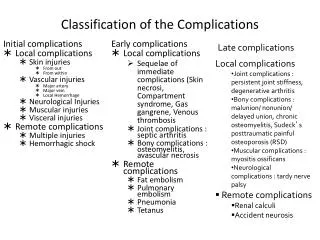
Classification of the Complications. Late complications. Initial complications Local complications Skin injuries From out From within Vascular injuries Major artery Major vein Local Hemorrhage Neurological Injuries Muscular injuries Visceral injuries Remote complications

E N D
Classification of the Complications Late complications Initial complications • Local complications • Skin injuries • From out • From within • Vascular injuries • Major artery • Major vein • Local Hemorrhage • Neurological Injuries • Muscular injuries • Visceral injuries • Remote complications • Multiple injuries • Hemorrhagic shock Early complications • Local complications • Sequelae of immediate complications (Skin necrosi, Compartment syndrome, Gas gangrene, Venous thrombosis • Joint complications : septic arthritis • Bony complications : osteomyelitis, avascular necrosis • Remote complications • Fat embolism • Pulmonary embolism • Pneumonia • Tetanus • Local complications • Joint complications : persistent joint stiffness, degenerative arthritis • Bony complications : malunion/ nonunion/ delayed union, chronic osteomyelitis, Sudeck’s posttraumatic painful osteoporosis (RSD) • Muscular complications : myositisossificans • Neurological complications : tardy nerve palsy • Remote complications • Renal calculi • Accident neurosis Salter RB. Textbook of Disorders and Injury of Musculoskeletal System
Treatment by MethodPlate & Screw Advantages Disadvantages Extensive soft tissue dissection Limitation of early weight bearing & function Inability to correct significant shortening deformity • rigidity of fixation • versatility for various anatomic locations & situations (e.g., periarticular deformities) • correction of deformities under direct visualization • safety following failed or temporary external fixation
Intramedullary Nail • Particularly useful in the lower extremity the strength &load-sharing characteristics • Ideal for correction of diaphysealdeformities • The method may also be useful for deformities at the metaphyseal-diaphysealjunction. • Also excellent for osteopenic bone where screw purchase may be poor
Indirect vs Direct bone healing • Indirect: • Ensure mechanical strength while bone heal • With increasing stress callus grow stronger (Wolf law) • Direct: • there is a long period during which the bone depends entirely upon the metal implant for its integrity. • the implant diverts stress away from the boneosteoporotic and not recover fully until the metal is removed
Applied fixation principles in osteoporotic bone Relative instead of absolute stability • Indirect, functional, not anatomical reduction • Locked splinting with long plates or nails • Load distribution, no peak stresses • No interfragmentary compression • Secondary bone healing with callus formation • No mixture of principles and methods