Download
1 / 32
330 likes | 374 Vues
Explore the link between oxidative stress and neurodegeneration, focusing on ROS generation, antioxidant mechanisms, & impact on brain cells. Discover how oxidative stress affects DNA, protein, and lipid components.

E N D
Review Article Mechanism of Oxidative Stress in Neurodegeneration Sonia Gandhi and Andrey Y. Abramov Department of Molecular Neuroscience, UCL Institute of Neurology, Queen Square, London WC1N 3BG, UK Hindawi Publishing Corporation Oxidative Medicine and Cellular Longevity Volume 2012, Article ID 428010, 11 pages doi:10.1155/2012/428010 Nafith Abu Tarboush DDS, MSc, PhD natarboush@ju.edu.jo www.facebook.com/natarboush Oxidative Stress & Neurodegeneration
Oxidative Stress & Neurodegeneration • Oxidative stress is important in their etiology (association) • Aging has been established as the most important risk factor (AD & PD) • Aging: cumulative oxidative stress leads to mitochondrial mutations, mitochondrial dysfunction, & oxidative damage • Is oxidative stress a result of dysfunctional & dying neurons? or • Does oxidative stress itself cause the dysfunctionality/death of neurons? • How does a global event such as oxidative stress result in the selective neuronal vulnerability seen in most neurodegenerative diseases? • & finally, if oxidative stress is truly fundamental to pathogenesis, then will the use of antioxidant therapy be successful?
OUTLINE • In order to address these questions: • Definition of oxidative stress • Show how ROS is generated in the human brain • The antioxidant defense mechanisms • Is there an evidence that oxidative stress can be found in neurodegenerative disease? • Is oxidative stress truly pathogenic in disease models? • What treatment experimental studies have been performed?
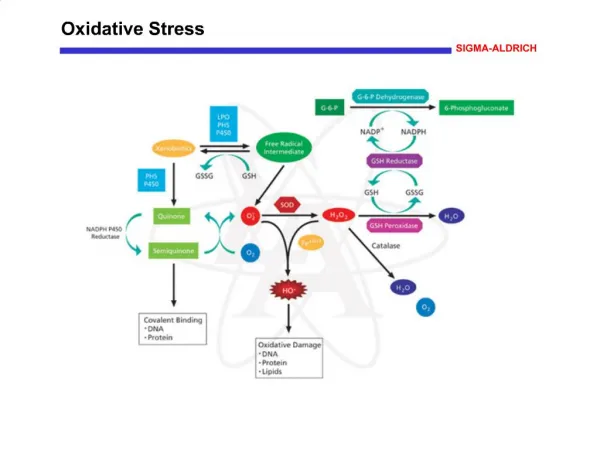
Oxygen, Brain & Oxidative Stress • Oxygen is essential for the normal function (respiration, high redox potential, excellent oxidizing agent) • Neurons & astrocytes, are responsible for the massive consumption of O2 (∼2% vs. >20%) • The state of hyperoxia produces toxicity (including neurotoxicity) • Partially reduced forms of oxygen are highly active (ROS) • Varieties of (ROS): superoxide (O•−2), hydrogen peroxide (H2O2), & hydroxyl radical (OH•) (the most reactive) • The modern use: radicals & non-radicals (O3, O2, OH–) • What do they do? Chemically interact with biological molecules • Aerobic organisms survive its presence only because they contain antioxidant defenses
Oxygen, Brain & Oxidative Stress • Brain cells require more effective antioxidant protection: • They exhibit higher (10-fold) oxygen consumption • Non-dividing cells (long life duration) • Nitric oxide has a prominent role in the brain (RNS) • Oxidative stress: is a condition in which the balance between production of ROS & level of antioxidants is significantly disturbed & results in damage to cells by excessive ROS • ROS may target several different substrates in the cell, causing protein, DNA, RNA oxidation, or lipid peroxidation
Oxygen, Brain & Oxidative Stress • Lipid peroxidation products of polyunsaturated fatty acids: especially arachidonic acid & docosahexanoic acid (DHA) which are abundant in brain, are malondialdehyde & 4-hydroxynonenal • ROS attacks protein, oxidizing both the backbone & the side chain, which in turn reacts with amino acid side chains to form carbonyl functions (oxidation can yield aldehydes and ketones) • ROS attacks nucleic acids in a number of ways, causing DNA-protein crosslinks, breaks in the strand, & modifies purine & pyrimidine bases resulting in DNA mutations
NADPH Oxidase • A multi-subunit enzyme complex • Is a member of the NOX gene family • Also called phagocytic oxidase (PHOX) • Seven NOX genes have been identified • The most expressed of the NOX enzymes in the brain is NOX2 • The enzyme transfers the proton across the membrane, & the end product of the enzyme is superoxide
Xanthine Oxidase • It is a molybdo-flavo-enzyme complex • A key enzyme of purine catabolism • XO catalyses the oxidation of a wide range of substrates & pass electrons to molecular oxygen to produce uric acid, superoxide, & hydrogen peroxide
Mitochondria • Mitochondria (electron transport chain-ETC), in contrast to other cellular producers of ROS, generate free radicals all the time • Mitochondria, which harbor the bulk of oxidative pathways, leak single electrons to oxygen • Depending on the metabolic conditions, isolated mitochondria produces superoxide in e.x.; • Respiratory complex I • Complex III • Aconitase • α-ketoglutarate dehydrogenase complex • The production of superoxide is dependent on the value of mitochondrial membrane potential
Mitochondria... Cont. • Inhibition of neuronal respiration leads to a significant increase in ROS in mitochondria • Overproduction of ROS in mitochondria leads to imbalance & induce oxidative stress & neurodegeneration • This effect can be reduced by mitochondrial un-couplers (how?) • Significant neuroprotection by mild uncoupling with UCP2 in cerebral stroke • Mutations in mitochondrial complexes I–IV leads to activation of ROS production & neuronal cell death
Monoamine Oxidase • Flavoenzymes • Mitochondrially located (outer membrane) • Monoamine oxidase A & B (MAO A & B); ∼70% identical • Their role in oxidative catabolism of important amine neurotransmitters (serotonin, dopamine, & epinephrine) • Expressed in neurons (MAO-A) & glial cells (MAO A & B) • MAO breaks down monoamines using FAD & results in the production of aldehydes. The FAD-FADH2 cycle generates hydrogen peroxide
Superoxide Dismutases • Play a crucial role in scavenging O•−2 • Specialized in eliminating superoxide anion radicals • Three distinct isoforms: • Copper-zinc superoxide dismutase (Cu/Zn SOD) • Manganese superoxide dismutase (Mn SOD) • Extracellular superoxide dismutase (EC SOD)
Glutathione Peroxidases • A family of multiple isozymes • Catalyze the reduction of H2O2 to water using reduced glutathione (GSH) as an electron donor • (H2O2 + 2GSH → GS-SG + 2H2O) • In mammalian tissues, there are four major selenium-dependent glutathione peroxidases • GPX1 is known to localize primarily in glial cells, in which GPX activity is 10-fold higher than in neurons
Catalase • Catalase is a ferriheme-containing enzyme • Converts hydrogen peroxide to water • It is localized in peroxisomes, cytoplasm & mitochondria
GSH • The main antioxidant in CNS • The most abundant small molecule, non-protein thiol in cells • Consists of a tripeptide • Reduced GSH can non-enzymatically act directly with free radicals, notably superoxide radicals, hydroxyl radicals, nitric oxide, & carbon radicals for their removal • GSH peroxidase & GSH reductase can act enzymatically to remove H2O2 & maintain GSH in a reduced state
Vitamin E • A lipid soluble molecule with antioxidant function (mainly) • It appears to neutralize the effect of peroxide & prevent lipid peroxidation in membranes
Alzheimer’s disease • The most common neurodegenerative disease, affecting approximately 16 million people worldwide • Characterized by progressive neuronal loss associated with aggregation of protein as extracellular amyloid (βA) plaques, & intracellular tau tangles • AD brains also show evidence of ROS mediated-injury; • Increase in levels of malondyaldehyde & 4-hydroxynonenal in brain & cerebrospinal fluid • Protein carbonyl moieties are increased in the frontal & parietal cortices, & hippocampus with sparing of the cerebellum • Increase in hydroxylated guanosine
Parkinson’s disease • The second most common • Characterized by progressive loss of dopaminergic neurons in the substantia nigra, & aggregation of the protein α-synuclein • Concentration of PUFAs in the substantia nigra is reduced, while the levels of lipid peroxidation markers (malondialdehyde & 4-hydroxynonenal) are increased • Protein oxidative damage in the form of protein carbonyls is also evident • Increased levels of 8-hydroxydeoxygua-nosine
Mechanisms of Oxidative Stress: ROS Production by Mitochondrial Dysfunction • Mitochondrial pathology is evident in many neurodegenerative diseases including AD & PD • The spectrum of mitochondrial dysfunction is vast; • Respiratory chain dysfunction • Oxidative stress • Reduced ATP production • Calcium dysregulation • Mitochondrial permeability transition pore opening • Deregulated mitochondrial clearance (mitophagy)
ROS Production by Mitochondrial Dysfunction; PD • A reduction in complex I activity in the substantianigra • The neurotoxin 1-methyl-4-phenyl-l,2,3,6-tetrahydropyridine (MPTP) has been shown to produce parkinsonian symptoms • l-methyl-4-phenylpyridinium (MPP+), the active metabolite of MPTP, can block ETC (same site as rotenone) • Rotenone or MPP+ also produces superoxide anions in sub-mitochondrial particles • Mild uncoupling of mitochondria with UCP2 overexpression reduces ROS production (MPP+, rotenone) • The identification of a number of PD-related genes that are strongly associated with mitochondrial function (PINK1, DJ-1, & Parkin)
oxidative stress is a primary event in PD pathogenesis • Mutations in PINK1 (mitochondrial kinase) cause a recessive form of PD • PINK1 deficiency results in inhibition of complex I, & rotenone-like increased production of ROS in mitochondria • Abnormal aggregation of protein α-synuclein, which accumulates in all PD brain • Mutations in α-synuclein gene cause a familial form of autosomal dominant PD • Expression of mutant α-synucleinin neurons results in increased ROS production • α-synuclein binds mitochondria & induce mitochondrial fragmentation
ROS Production by Mitochondrial Dysfunction; AD • A reduction in complex IV activity in mitochondria from the hippocampus • Deregulation of calcium homeostasis; • βA causes increased cytoplasmic calcium levels & mitochondrial calcium overload, resulting in increase in ROS production & opening of the PTP • βA directly interact with cyclophilin D (a PTP component) forming a complex in the mitochondria that has reduced threshold for opening • Fragmented mitochondria are seen in AD hippocampus
Use of Antioxidant Therapy in Neurodegenerative Disease • The rationale for the use of antioxidants as therapies is clear • The benefits of antioxidants in animal & cell models of disease was promising • Vitamin E • Vitamin C • Coenzyme Q
Promising ! • Vitamin E supplementation in AD mouse model resulted in improved cognition & reduced βA deposition • AD; Daily injections of vitamin C in mouse model significantly reduced memory deficits • PD; Coenzyme Q has been shown to have multiple protective effects within the mitochondria • PD;CoQprotects MPTP-treated mice from dopaminergic neuronal loss & also attenuated α-synuclein aggregation
Promising but! • There has been no proven benefit for the use of vitamin E &/or vitamin C in either AD or PD from large randomised controlled clinical trials • Vitamin E, CoQ, & glutathione clinical trials in PD concluded that there were only minor treatment benefits in the CoQ trials that may have been due to improvement in the respiratory chain deficit rather than a direct antioxidant action • None of the trials have shown significant benefit to warrant recommendation for use in the clinical setting!!!!
Promising but! • All animal models are limited in recreating the human disease • long-time frame • Gradual accumulation of age-related changes • Antioxidants must be administered at an early stage where the process influences pathogenesis most • The bioavailability of reducing molecules in the human brain in the doses used in animal models • The effective targeting of such molecules to the mitochondria in human brain • Several different producers of oxidative stress in each disease (need to be targeted separately but simultaneously)