Download
1 / 62
720 likes | 1.75k Vues
Fundamental Nursing Chapter 29 Gastrointestinal Intubation.

E N D
Clients, especially those undergoing abdominal or gastrointestinal (GI) surgery, may require some type of tube placed within their stomach or intestine. Use of a gastric or intestinal tube reduces or eliminates problems associated with surgery or conditions affecting the GI tract such as impaired peristalsis, vomiting, or gas accumulation. Tubes also can nourish clients who cannot eat. • This chapter discusses the multiple uses for gastric and intestinal tubes and the nursing guidelines and skills for managing associated client care.
Intubation • Intubation generally means the placement of a tube into a body structure; in this chapter, it refers specifically to insertion of a tube into the stomach or intestine by way of the mouth or nose.
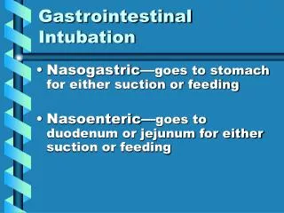
Orogastric intubation (insertion of a tube through the mouth into the stomach), nasogastric intubation (insertion of a tube through the nose into the stomach; Fig. 29-1), and nasointestinal intubation (insertion of a tube through the nose to the intestine) are performed to remove gas or fluids or to administer liquid nourishment.
A tube also may be inserted within an ostomy (surgically created opening). A prefix identifies the anatomic site of the ostomy; for instance, a “gastrostomy” is an artificial opening into the stomach
Gastric or intestinal tubes are used for a variety of reasons, including the following: • Performing a gavage (providing nourishment) • Administering oral medications that the client cannot swallow • Obtaining a sample of secretions for diagnostic testing • Performing a lavage (removing substances from the stomach, typically poisons) • Promoting decompression (removing gas and liquid contents from the stomach or bowel) • Controlling gastric bleeding, a process called compression or tamponade (pressure)
Types of Tubes • Although all gastric and intestinal tubes have a proximal and distal end, their size, construction, and composition vary according to their use (Table 29-1). • Tubes can be identified according to the location of their insertion (mouth, nose, or abdomen) or the location of their distal end (stomach [gastric] or intestinal).
1. Orogastric Tubes • An orogastric tube (tube inserted at the mouth into the stomach), such as an Ewald tube, is used in an emergency to remove toxic substances that have been ingested. The diameter of the tube is large enough to remove pill fragments and stomach debris
2. Nasogastric Tubes • A nasogastric tube (tube placed through the nose and advanced to the stomach) is smaller in diameter than an orogastric tube but larger and shorter than a nasointestinal tube. Some nasogastric tubes have more than one lumen (channel) within the tube. with multiple uses: decompression to remove fluid and gas from the stomach
Because nasogastric tubes remain in place for several days or more, many clients complain of nose and throat discomfort. • Furthermore, gastric tubes tend to dilate the esophageal sphincter, • The stretched opening may contribute to gastric reflux (reverse flow of gastric contents), If gastric reflux occurs, the liquid could enter the airway and interfere with respiratory function.
3. Nasointestinal Tubes • Nasointestinal tubes (tubes inserted through the nose for distal placement below the stomach) are longer than their gastric counterparts. • They are used to provide nourishment (feeding tubes) or to remove gas and liquid contents from the small intestine (decompression tubes).
4. Transabdominal Tubes • Transabdominal tubes (tubes placed through the abdominal wall) provide access to various parts of the GI tract. Two examples are a gastrostomy tube or G-tube (transabdominal tube located within the stomach) • A gastrostomy tube is placed surgically or with the use of an endoscope. (Fig. 29-4A).
Figure 29-4 • Transabdominal tubes. ( A) Percutaneous endoscopic gastrostomy (PEG) tube. (B) Percutaneous endoscopic jejunostomy (PEJ) tube
Nasogastric Tube Management • Usually nurses insert nasogastric tubes. Additional nursing responsibilities include keeping the tube patent (or unobstructed), implementing the prescribed use, and removing the tube when it has accomplished its therapeutic purpose.
Insertion • Inserting a nasogastric tube involves preparing the client, conducting preintubation assessments, and placing the tube.
Client Preparation • Most clients are anxious about having to swallow a tube. • Explaining the procedure and giving instructions on how the client can assist while the tube is being passed may further reduce anxiety.
Preintubation Assessment • Level of consciousness • Weight • Bowel sounds • Abdominal distention • Integrity of nasal and oral mucosa • Ability to swallow, cough, and gag • Any nausea and vomiting
One main goal of the assessment is to determine which nostril is best to use when inserting the tube and the length to which the tube will be inserted.
Nasal Inspection • the nurse inspects each nostril for size, shape, and patency. The client should exhale while each nostril in turn is occluded. The presence of nasal polyps (small growths of tissue), a deviated septum (nasal cartilage deflected from the midline of the nose), or a narrow nasal passage excludes a nostril for tube insertion.
Tube Measurement • before inserting a tube, the nurse obtains the client's NEX measurement (length from nose to earlobe to the xiphoid process [tip of the sternum]; Fig. 29-5) and marks the tube appropriately.
The first mark on the tube is made at the measured distance from the nose to the earlobe. It indicates the distance to the nasal pharynx, a location that places the tip at the back of the throat but above where the gag reflex is stimulated. A second mark is made at the point where the tube reaches the xiphoid process, indicating the depth required to reach the stomach.
Tube Placement • When inserting a nasogastric tube, the nurse's primary concerns are to cause as little discomfort as possible, to preserve the integrity of the nasal tissue, and to locate the tube within the stomach, not in the respiratory passages. • Once the tube is at its final mark, the nurse must verify the location within the stomach.
The physical assessment methods that nurses use to determine the distal location of a nasogastric tube are as follows: • Aspirating fluid: If aspirated fluid appears clear, brownish-yellow, or green, the nurse can presume that its source is the stomach (Fig. 29-6). • Auscultating the abdomen: The nurse instills 10 mL or more of air while listening with a stethoscope over the abdomen. If a swooshing sound is heard, the nurse can infer that the cause was air entering the stomach. Belching often indicates that the tip is still in the esophagus.
Testing the pH of aspirated liquid: The first two techniques provide only presumptive signs that the tube is in the stomach; testing pH confirms acidic gastric contents. Other than obtaining an abdominal x-ray, the pH test is the most accurate technique for checking tube placement. See Nursing Guidelines 29-1
Once the nurse has confirmed stomach placement (using two methods is best), he or she secures the tube to avoid upward or downward migration (Fig. 29-8).
Figure 29-8 • (A) One end of a piece of tape is split, forming two narrower strips, and the opposite end is left intact. (B) The wider intact end of the tape is applied to the nose, and the narrower strips are wound around the tube in opposite directions to secure the nasogastric tube.
Use and Maintenance • Nasogastric tubes are connected to suction for gastric decompression or are used for tube feeding.
Gastric Decompression • Suction is either continuous or intermittent. • The tube is connected to a wall outlet or portable suction machine (Fig. 29-9).
Figure 29-9 • Suction removes liquids and gas from the stomach.
Promoting Patencywith intermittent suctioning • Giving ice chips or occasional sips of water to a client who is otherwise NPO promotes tube patency. The fluid helps to dilute the gastric secretions.
Restoring Patency • The nurse assesses tube patency frequently by monitoring the volume and characteristics of drainage and observing for signs and symptoms suggesting an obstruction (nausea, vomiting, and abdominal distention). • Sometimes the nasogastric tube must be irrigated to maintain or restore patency (Skill 29-2).
Removal • Nurses remove a nasogastric tube (Skill 29-3) when the client's condition improves, when the tube becomes hopelessly obstructed, or according to the agency's standards for maintaining the integrity of the nasal mucosa.
Unobstructed larger-diameter tubes usually are removed and changed at least every 2 to 4 weeks for adults. Small-diameter, flexible tubes are removed and changed every 4 weeks to 3 months, depending on agency policy.
Transabdominal Tube Management • The physician inserts transabdominal tubes, such as gastrostomy and jejunostomy tubes, but the nurse is responsible for assessing and caring for them and their insertion sites. Conscientious care is necessary because gastrostomy tubes may leak (Box 29-1) and cause skin breakdown. See Nursing Guidelines 29-3.
Figure 29-12 • Inspection. (A) Inspecting for drainage. (B) Inspecting the skin.
Box 29-1 • Causes of Gastrostomy Leaks • Disconnection between the feeding delivery tube and G-tube • Clamped G-tube while tube feeding is infusing • Mismatch between the size of the G-tube and stoma • Increased abdominal pressure from formula accumulation, retching, sneezing, coughing • Underinflation of the balloon beneath the skin • Less than optimal stoma or stomal location
Providing nutrition by the oral route is always best. However, if oral feedings are impossible, nourishment is provided enterally or parenterally (see Total Parenteral Nutrition, Chap. 16). • Tube feedings are used when clients have an intact stomach or intestinal function but are unconscious, have undergone extensive mouth surgery, have difficulty swallowing, or have esophageal or gastric disorders.
Benefits and Risks • For example,dumping syndrome (cluster of symptoms from the rapid deposition of calorie-dense nourishment into the small intestine). The symptoms, which include weakness, dizziness, sweating, and nausea, are caused by fluid shifts from the circulating blood to the intestine and low blood glucose level. Diarrhea also may result when administering hypertonic formula solutions.
Formula Considerations • In addition to the type of tube and the access site, the type of formula also is individualized, based on the client's nutritional needs (Table 29-4). Factors include the client's weight, nutritional status, and concurrent medical conditions and the projected length of therapy.
Tube-Feeding Schedules • Tube feedings may be administered on bolus, intermittent, cyclic, or continuous schedules.
Bolus Feedings • A bolus feeding (instillation of liquid nourishment in less than 30 minutes four to six times a day) usually involves 250 to 400 mL of formula per administration.
Intermittent Feedings • An intermittent feeding (gradual instillation of liquid nourishment four to six times a day) is administered over 30 to 60 minutes, the time most people spend eating a meal. The usual volume is 250 to 400 mL per administration.