Download
1 / 40
440 likes | 973 Vues
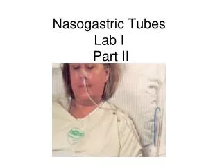
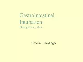
Gastrointestinal Intubation Nasogastric tubes. Enteral Feedings. Objectives. To know the types of nasogastric tubes To understand the indications for their use To know the technique of insertion of NG tubes To know the complications of NG tubes

E N D
Gastrointestinal IntubationNasogastric tubes Enteral Feedings
Objectives • To know the types of nasogastric tubes • To understand the indications for their use • To know the technique of insertion of NG tubes • To know the complications of NG tubes • To understand the meaning of enteral Feedings • To know the indications and Complications. • To know the meaning of gastrostomy
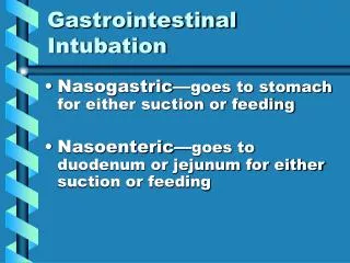
Nasogastric tube • Gastrointestinal intubation is inserting of rubber or plastic tube into the stomach , duodenum or intestinal • The tube inserted through mouth .nose , or abdominal ( gastrostomy .jejunostomy )
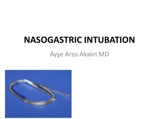
Types of Tubes • Short tubes: passed through the nose into the stomach • Levin tube: range in size from 14 to 18 Fr, single lumen made of plastic or rubber with holes near the tip. • Gastric Sump(Salem) : is radiopaque, clear plastic double lumen
Types Cont. • Medium Tubes: tubes are passed through the nose to the duodenum and the jejunum. Used for feeding • Polyurethane or silicone rubber feeding tubes have a narrower diameter (6 to 12fr) and require the use of a stylet for insertion • Long tubes: passed through the nose, through the esophagus and stomach into the intestines. Used for decompression of the intestines.
Indications for GI Intubation • To decompress the stomach and remove gas and fluid • To lavage the stomach and remove ingested toxins • To diagnose disorders of GI motility and other disorders • To administer medications and feedings • To treat an obstruction • To compress a bleeding site • To aspirate gastric contents for analysis
Intubating the client with an NG tube • Assessment: • Who needs an NG: • Surgical clients • Ventilated client • Neuromuscular impairment . • Clients who are unable to maintain adequate oral intake to meet metabolic demands. • Assess patency of nares.
Assessment cont. • Assess client’s medical history: • Nosebleeds • Nasal surgery • Deviated septum • Anticoagulation therapy • Assess client’s gag reflex. • Assess client’s mental status. • Assess bowel sounds.
Technique • Gather equipment: • 14 0r 16 Fr NG tube • Lubricating jelly • PH test strips • Tongue blade • Flashlight • Emesis basin • syringe • 1 inch wide tape or commercial fixation device • Suctioning available and ready
Technique contu. • Explain procedure to client • Position the client in a sitting or high fowlers position. If comatose-semi fowlers. • Examine feeding tube for flaws. • Determine the length of tube to be inserted. • Measure distance from the tip of the nose to the earlobe and to the xyphoid process of the sternum. • Prepare NG tube for insertion.
Fowler's Position. Used to promote drainage or ease breathing. Head rest is adjusted to desired height and bed is raised slightly under patient's knees
Implementation • Wash Hands • Put on clean gloves • Lubricate the tube • Hand the client a glass of water • Gently insert tube through nostril to back of throat (posterior naso pharynx). Have client flex head toward chest after tube has passed through naso pharynx
Implementation Cont. 6)Emphasize the need to mouth breathe and swallow during the procedure. 7) Swallowing facilitates the passage of the tube through the oropharynx. 8) When the tip of the tube reaches the carina stop and listen for air exchange from the distal end of the tube. If air is heard remove the tube. 9) Advance tube each time client swallows until desired length has been reached. 10) Do not force tube. If resistance is met or client starts to cough, choke or become cyanotic stop advancing the tube and pull back.
Implenentation Cont. 11) Check placement of the tube. X-ray confirmation Testing pH of aspirate inject air and auscultate it 12) Secure the tube with tape or commercial device
Evaluation • Observe client to determine response to procedure. • ALERTS!!! Persistent gagging – prolonged intubation and stimulation of the gag reflex can result in vomiting and aspiration • Coughing may indicate presence of tube in the airway.
Evaluation Cont. • Note location of external site marking on the tube • Documentation • Size of tube, which nostril and client’s response. • Record length of tube from the nostril to end of tube • Record aspirate pH and characteristics
Testing Placement • Wash hands and put on clean gloves • Draw up 30cc of air into the syringe and attach to end of the NG tube. Flush tube with 30cc of air prior to attempting to aspirate fluid. Draw back on the syringe to obtain 5 to 10 cc of gastric aspirate. • If unable to aspirate: • Advance tube – may be in air space above aspirate level • If intestinal placement suspected (pH 4-6) withdraw tube 5 to 10 cm • Have client lie on his/her left side wait 10-15 mins and attempt aspiration again.
Testing Placement cont. • Observe appearance of aspirate: • From client with enteral feeding – appearance of curdled enteral feed • From nasointestinal – bile stained • From stomach (non feed)– green, tan, bloody, brown. • Pleural fluid – pale yellow and serous • Gently mix aspirate in syringe
Testing Placement Cont. Measure pH of aspirated GI contents by dipping pH strip into the fluid or by applying a few drops of the fluid to the strip. Compare the color of the strip with the color on the chart. • Gastric fluid from a client who has fasted for at least 4 hours usually has a pH range from 1 to 4 but may be increased if the client is receiving acid inhibiting medications (pH 4-6)
Testing Placement Cont. • Fluid from nasointestinal tube of fasting client usually has a pH greater than 6. intestinal contents are less acidic than stomach. • Clients with a continuous tube feed may have a pH of 5 or higher. • Pleural fluid from the tracheubronchial tree is generally greater than 7. • National Patient Safety Association(2005a) recommend a pH of less than 5.5 feedings can be initiated (Best, 2005)
Testing Placement Cont. • Measure the length of the tube from nostril to tip. • If after repeated attempts, it is not possible to aspirate fluid from a tube that was originally established by x-ray examination to be in the desired position and there are NO risk factors for dislocation, tube has remained in original position and the client is NOT experiencing any difficulty the nurse may assume the tube is correctly placed.
Enteral Nutrition • What is it: • The administration of nutrients directly into the GI tract. The most desirable and appropriate method of providing nutrition is the oral route, but this is not always possible. • Nasogastric feeding is the most common route • Nurses are the main healthcare professional responsible for intubation
Administering Enteral Feeds • Indications: • Clients who are unable to maintain adequate oral intake to met metabolic demands • Surgical cases • Ventilated clients • Neuromuscular impairment • Clients requiring bowel rest. • Generally these clients have been referred to the Dietician.
Administering Enteral Feeds • Contraindications: • Clients with diffuse peritonitis. • Severe pancreatitis • Intestinal obstruction • Severe D&V • Paralytic ileus.
Complications • Clogged Tube- most common • Flush tube with 30-60 cc q4h if continues feed. Use liquid meds when possible. Flush tube after giving medication. • Dumping Syndrome: solution with high osmolality- water moves into stomach and intestines from the fluid surrounding the organs and vascular system causing dehydration, hypotension and tachycardia • Aspiration : ensure head of bed is elevated at least 30 degrees while feeds are being administered
Complications Cont. • Dehydration- diarrhea is a common problem. • Electrolyte imbalance: hyperkalemia and hypernatremia • Oral mucosal breakdown • Nasal irritation
Gastrostomy • Surgical procedure in which an opening is created into the stomach • Preferred route for prolonged nutrition((greater than 3 to 4 weeks) • Preferred in clients who are comatose – decreases the risk for regurgitation and aspiration
Methods of Insertion • Percutaneous endoscopic gastrostomy (PEG) may be clamped between feedings • Low-profile gastrostomy device (LPGD) may be inserted 3-6 months after initial gastronomy tube placement
Client Education • Clients can go home and administer their own feeds, (or caregiver ) • Educational needs: • Teach how to administer a bolus feed • How to assess residual volumes before feeds • How to maintain patency of tube with flushing of tube pre and post feeds and medications • Elevating head of bed while feeds are administered and 1 hour following • Monitor tube length