Download
1 / 21
210 likes | 417 Vues
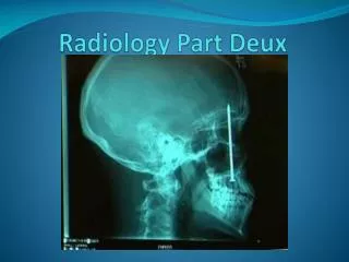
Radiology Part Deux. CT Scans (CAT Scans). CT scanning or ( CAT scanning ) is using X-rays to create a 3D image of the inside of an object. CT stands for computed tomography . Tomography is developing an image in sections or slices. History.

E N D
CT Scans (CAT Scans) • CT scanning or (CAT scanning) is using X-rays to create a 3D image of the inside of an object. • CT stands for computed tomography. • Tomography is developing an image in sections or slices.
History • Believe it or not, the first slice of tissue photographed on x-ray film (tomography) was done by Alessandro Vallebona in the early 1900s.
History • When computers developed and technology advanced, two men, the Brit, Godfrey Hounsfield and the South African Allan Cormack won the Nobel Prize in Medicine in 1979 for creating the first CT scanner independently. • The funding of the mass production of these machines was done by the same company that produced Beatles albums, EMI! First scanner funded by EMI
CT Scan Usage • CT scans are the “gold standard” in the diagnosis of a large number of diseases. • More recently they have been used in preventative medicine, screening patients at high risk before they develop the condition.
Main Items CT Scans are used for • Brain injuries, bleeding in the brain and skull fractures.
Main Items CT Scans are used for Brain tumors.
Main Items CT Scans are used for Stroke patients and aneurysm patients in tracking blood vessel leakage.
Main Items CT Scans are used for Detection of airspace disease in the lungs (i.e. emphysema).
Main Items CT Scans are used for Tumors in the colon and other obstructive bowel conditions.
Procedure for a CT Scan • Patient, depending on the problematic area, may need to ingest a radioactive dye to better show up the body regions during the scan. • An X-ray rotates around the body or body part and takes pictures from all angles. • The pictures are pieced together on a computer screen to give a 3D image of the affected area.
Cons against CT Scans • The major knock against CT scans is the cost. • Other people argue about the dosage of X-rays a patient takes in from each scan are too high. • Lots of tests are not conclusive, so other imaging techniques or exploratory surgery may still be needed.
Magnetic Resonance Imaging (MRI) • An MRI is used to image the internal structures of the body. • It is done with much better clarity than a CT scan. • It is useful in cancer detection, joint injuries, brain injuries and cardiovascular problems.
MRI • An MRI uses no radiation, but instead uses a power magnetic field to align all the hydrogen atoms in the water molecules of the body. • Radio waves are then used to create an image based on the alignment of the hydrogen atoms.
History • MRI is a newer technology, having first been used in the early 1970s. • They are now more widespread, although they are just as expensive to build and maintain as CT scans. • Paul Lauterbur of the University of Illinois, just won a Nobel Prize in 2003 for his work on improving MRIs.
MRI Procedure • Very similar to a CT scans, however no dye needs to be ingested. • The MRI machine passes over the patient and uses radio waves and a high powered magnetic field to create a 3D image.
MRI vs. CT Scans MRI CT Scans • Expensive • Better resolution at distinguishing a contrast between two similar looking tissues. • MRI can image in any plane. • Due to the magnetic field, certain problems can arise with implants (pacemakers, metal plates, hearing aids, etc…). No radiation problems though. • Expensive • Better resolution at determining individual tissues. • CT scans usually can only image along the axial plane of the body (from head to toe). • No real problems with implants, just the amount of radiation.
PET Scans • Positron emission tomography is another form of medical imaging. • It is used for imaging tumors, tracing blood flow and looking at brain activity to determine brain disease/dementia.
How PET works • The patient is injected with a radioactive “tracer” molecule. • Wait until the tracer gets incorporated into the tissue in question. • Send the patient through the PET scanner which gets a picture from the gamma ray radiation given off by the tracer molecules in the tissue.
PET Info • It was developed at Massachusetts General Hospital in the 1950s. • It is rare to be used on its own. Usually used to confirm information given by a CT scan or MRI. • It uses radioisotopes, which give off radiation, so there is a risk of radiation exposure.
Homework • Do you think the different radiology options are cost feasible? Why or why not? • Rank CT Scans, MRIs and PET scans in terms of safety. Why did you rank them as you did? • Compare CT scans, MRIs and PET scans in terms of what they can best diagnose. Which technology do you feel would be the best for the following: • Cracked skull • Bleeding in the brain • Alzheimer's detection • Torn elbow ligaments