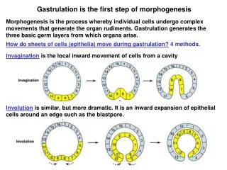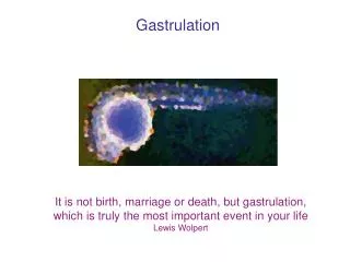
Gastrulation
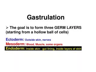
Gastrulation. The goal is to form three GERM LAYERS (starting from a hollow ball of cells) Ectoderm: Outside skin, nerves Mesoderm: Blood, Muscle, some organs Endoderm: Inside skin- -gut lining, inside layers of skin. Gastrulation involves changes in cell shape

Gastrulation
E N D
Presentation Transcript
Gastrulation • The goal is to form three GERM LAYERS (starting from a hollow ball of cells) • Ectoderm: Outside skin, nerves • Mesoderm: Blood, Muscle, some organs • Endoderm: Inside skin- -gut lining, inside layers of skin
Gastrulation involves changes in cell shape and changes in cell adhesion
Cytoskeletal events drive cell shape changes Contraction of the adhesion belt drives apical constriction (see Alberts Fig 20-26)
21_24_Adherens_junct.jpg Alberts Fig. 20-25
21_21_cell_cell_junction.jpg E-cadherin Alberts Fig. 20-22
Types of Movement in Gastrulation Groups of cells Individual cells Local inward buckling of an epithelium Inward movement of a cell layer around a point or edge Movement of individual cells or small groups from an epithelium into a cavity Migration Movement of individual cells over other cells or matrix Splitting layers of cells (sometimes used to describe coordinated ingression) Spread of an outside cell layer (as a unit) to envelop a yolk mass or deeper layer Fig. 5.4
More complex changes in cell shape can drive elongation or shortening of a flat sheet of cells 15 cells “Convergent Extension” 4 cells Cell intercalation Narrowed and lengthened sheet of cells 30 cells 2 cells
Sea urchin gastrulation Our “simple” model Fig. 5.14 blastocoel
Sea urchin gastrulation Our “simple” model
Step 1: Primary mesenchyme cells ingress Inside Outside (apical) Mesenchyme cells- cells that are unconnected to one another and operate as independent units See also Figure 5.16
Primary mesenchyme ingression is driven by changes in cell adhesion Figure 5.16
Changes in cell adhesion drive the first step of gastrulation basal lamina and extracellular matrix
Invaginating primary mesenchyme cells beginning to migrate on the extracellular matrix lining the blastocoel
Primary mesenchyme cells migrate along the extracellular matrix using filopodia to detect chemical cues
Primary mesenchyme cells eventually fuse and form the spicules (skeletal rods) Figure 5.15 Figure 5.17
Step 2: Apical constriction and changes in the extracellular matrix create a dome-shaped invagination = archenteron (primitive gut) blastopore = opening Figure 5.19
Invagination of the Vegetal Plate involves changes in the extracellular matrix (CSPG)
Step 3: Cell intercalation (convergent extension) converts the dome (archenteron) into an elongated tube Figure 5.20
Step 4: Secondary mesenchyme cells at the leading edge of the gut tube use filopodia to look for cues at the animal pole and pull themselves to that site Ectoderm These secondary mesenchyme cells will become muscle (mesoderm) Figure 5.21 Endoderm (gut)
Pluteus larva Pluteus larva Figure 5.14
Sea urchin Early cleavage in Xenopus animal vegetal Fig. 7.2 Here is where gastrulation starts
Early cleavage in Xenopus animal vegetal Two functions of the blastocoel: 1. Prevents cells from interacting too soon 2. allows space for cell migrations during gastrulation
Sea urchin A Fate Map of the Xenopus Blastula Most Exterior Cells form ectoderm or endoderm Most Interior Cells form mesoderm Fig. 7.5 Mesoderm
Frog gastrulation: added complexity but similar mechanisms 1. Blastopore Formation sperm entry (That looks familiar!) Fig. 7.6
Mechanism #1 Apical constriction of bottle cells drives blastopore invagination Figure 7.7 Archenteron
Frog gastrulation: added complexity but similar mechanisms 2. Involution of Marginal zone cells Mechanism #2 INVOLUTION around dorsal lip Marginal Zone Cells inside MZ Fig. 7.6 outside MZ
Types of Movement in Gastrulation Local inward buckling of an epithelium Inward movement of a cell layer around a point or edge Movement of individual cells or small groups from an epithelium into a cavity MIGRATION Movement of individual cells over other cells or matrix Figure 5.4 Splitting layers of cells (sometimes used to describe coordinated ingression) Spread of an outside cell layer (as a unit) to envelop a yolk mass or deeper layer
2. Involution of marginal zone cells • movement ofinside MZ cellsdependent on ectoderm cells of blastocoel roof secreting fibronectin inside MZ Figure 10.7 outside MZ
Fibronectin is essential for mesodermal cell involution during gastrulation Control embryo Embryo injected with fibronectin competitor Yolk Plug Figure 7.12
3. Formation of the Archenteron = Convergent Extension of the Dorsal Mesoderm convergence and extension in three dimensions Figure 7.6
4. Epiboly of the Ectoderm Figure 7.6
Types of Movement in Gastrulation Local inward buckling of an epithelium Inward movement of a cell layer around a point or edge Movement of individual cells or small groups from an epithelium into a cavity MIGRATION Movement of individual cells over other cells or matrix Splitting layers of cells (sometimes used to describe coordinated ingression) Spread of an outside cell layer (as a unit) to envelop a yolk mass or deeper layer Figure 5.4
4. Epiboly of the Ectoderm Figure 7.9
5. mesenchymemigration Just like sea urchin Figure 7.6
Types of Movement in Gastrulation Local inward buckling of an epithelium Inward movement of a cell layer around a point or edge Movement of individual cells or small groups from an epithelium into a cavity MIGRATION Movement of individual cells over other cells or matrix Splitting layers of cells (sometimes used to describe coordinated ingression) Spread of an outside cell layer (as a unit) to envelop a yolk mass or deeper layer Figure 5.4
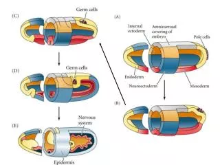
Gastrulation: Mission Accomplished Ectoderm Mesoderm Endoderm
Ectoderm (outer layer)will produce skin & the central nervous system (brain, spinal cord) through later invagination of the neural tube. In vertebrates, migrating neural crest cells form the peripheral nervous system & many other structures, including some bone, cartilage, and connective tissue in the head. Ectoderm
MESODERM (middle layer)will produce muscles, connective tissue, blood and blood vessels. In vertebrates also the notochord (progenitor of vertebrae), bones & cartilage, circulatory and urogenital systems (kidneys, gonads). Mesoderm
ENDODERM (inner layer)will produce the gut (entire digestive system) and other internal organs that arise as outpocketings of gut in vertebrates such as liver, lungs, pancreas, and salivary glands. Endoderm
Cleavage and Gastrulation Hatch from Zona Pellucida Fig. 8.20 Gastrulation Fig. 8.15
In mammals, gastrulation initiates AFTER formation of the placental connection to mom Fig. 8.23