Gastrulation and Neurulation
460 likes | 1.92k Vues
Gastrulation and Neurulation. 陳建榮. http://web.nchu.edu.tw/pweb/users/chenjr/. GASTRULATION 原腸形成 -Gastrulation : embryonic disc from bilaminar develops into trilamina -the embryo is so called gastrula . Ectoderm : epidermis, nervous system

Gastrulation and Neurulation
E N D
Presentation Transcript
Gastrulation and Neurulation 陳建榮 http://web.nchu.edu.tw/pweb/users/chenjr/
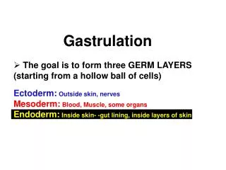
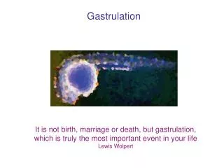
GASTRULATION原腸形成 • -Gastrulation: embryonic disc from bilaminar develops into trilamina • -the embryo is so called gastrula. • Ectoderm: epidermis, nervous system • Mesoderm: smooth muscular coats, connective tissue, and vessels; also blood cells and born marrow, skeleton, striated muscles, reproductive and excretory organs. • Endoderm: epithelial linings of respiratory passages and GI tract, including liver and pancreas Gastrulation
Primitive Streak原條 • At the beginning of the 3rd week, thickened linear band of epiblast on the dorsal caudal aspect of embryonic disc. • Primitive node原結(and primitive pit原凹) is on its cranial end. • Primitive groove原溝on the primitive streak. • Mesenchyme間葉細胞(from epiblast cells through primitive groove) • intraembryonic mesoderm胚內中胚層(mesoblast中胚葉) • intraembryonic endoderm胚內內胚層 • The process of forming mesoderm through primitive streak slows down after the end of the 2nd week and degenerates in the sacrococcygeal region of embryo.
Primitive Streak原條 • At the beginning of the 3rd week, thickened linear band of epiblast on the dorsal caudal aspect of embryonic disc. • Primitive node原結(and primitive pit原凹) is on its cranial end. • Primitive groove原溝on the primitive streak. • Mesenchyme間葉細胞(from epiblast cells through primitive groove) • intraembryonic mesoderm胚內中胚層(mesoblast中胚葉) • intraembryonic endoderm胚內內胚層 • The process of forming mesoderm through primitive streak slows down after the end of the 2nd week and degenerates in the sacrococcygeal region of embryo.
Prechordal plate脊索前板 oropharyngeal membrane口咽膜 mouth Cloacal membrane泄殖腔膜 anus
Cardiogenic area Notochordal process Prechordal plate Trilaminar embryonic disc 三層胚盤 Notochordal process 脊索突 Cardiogenic mesoderm 心臟生成中胚層 Cloacal membrane
Notochord脊索 • The primitive axis of the embryo and the future site of vertebral column • Formation of the Notochord • Fate of the Notochord • Degenerates as nucleus pulposus髓核 in the intervertebral disc • Notochord induces the development of neural plate
Formation of the Notochord: 1. Notochordal canal--extends from primitive pit and extend within notochordal process. Notochordal process extends from primitive node to prochordal plate. 2. Floor of notochordal process fused with underlying endoderm. 3. Opening of the floor of notochordal process. Notochordal canal communicates with yolk sac. 4. Notochordal canal disappears; primitive pit is now called neurenteric canal神經原腸管. The remain of notochordal process is flattened and called notochordal plate脊索板. 5. Notochordal plate infolds to form notochord脊索. 6. Notochord detaches from endoderm. Notochordal canal Endoderm
Notochord脊索 • The primitive axis of the embryo and the future site of vertebral column • Formation of the Notochord • Fate of the Notochord • Degenerates as nucleus pulposus髓核 in the intervertebral disc • Notochord induces the development of neural plate
Allantois尿囊 • It appears on day 16 from caudal end of yolk sac into connecting stalk. • It is involved with early blood formation and associated with urinary bladder development. • It becomes urachus臍尿管 and remains as median umbilical ligament正中臍韌帶. Yolk sac Connecting stalk umbilical cored
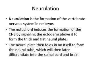
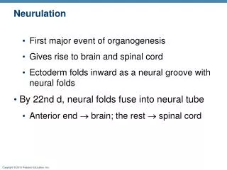
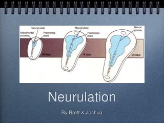
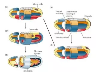
NEURULATION神經管形成: FORMATION OF NEURAL TUBE • The formation of neural plate and neural folds and the closure of these folds to form the neural tube. It is completed by the end of the 4th week. • The embryo is called neurula during neurulation. • Neural Plate神經板 and Neural Tube神經管 • Neuroectoderm--neural plate induced by notochord and develops into CNS • Neural plate ( notochord) - neural folds and neural groove • End of the 3rd week, neural folds neural tube. • Both ends of neural tube are cranial and caudal neuropores神經孔. • Neural Crest神經脊 Formation • -The crest of each neural fold detaches when neural tube forms. • -forms sensory ganglia of spinal (dorsal root ganglia) and cranial nerves (V, VII, IX, X). Also, its forms the Schwann cells, pia mater, arachnoid, pigment cells and adrenal medulla.
Secondary neurulation二次神經管形成 • - Neuropores close: • Cranial neuropore (day 24) forebrain • Caudal neuropore (day 26) somite 31(S2) • Mesodermal caudal eminence中胚層後隆起 neural cord neural tube Neural tube (CNS) and neural crest (PNS) derived from ectoderm Caudal neural tube derived form mesoderm
Sacrococcygeal teratoma 薦尾骨畸胎瘤
DEVELOPMENT OF SOMITES • - Both lateral sides of notochord mesoderm forms • - paraxial mesoderm軸旁中胚層 • -intermediatemesoderm中間中胚層 • - lateral mesoderm外側中胚層 • Somites • Somites appears first at occipital region at the end of the 3rd week • Give rise to axial skeleton (bone of head, neck and trunk), associated musculature and dermis of skin • # Somites are so prominent as to be a criteria for determining the age of embryo in 4th and 5th weeks.
E21 days E22 days E24 days
DEVELOPMENT OF THE INTRAEMBRYONIC COELOM • Intraembryonic coelom (cavity)胚內體腔--fusion of isolated coelomic spaces in the lateral mesoderm and cardiogenic mesoderm into a horseshoe-shaped cavity. • Lateral mesoderm divided into: • Parietal layer壁層--together with extraembryonic mesoderm covering amnion and ectoderm called somatopleure體壁, embryonic body wall (upper side) • Visceral layer臟層--together with extraembryonic mesoderm covering yolk sac and endoderm is called splanchnopleure臟壁, embryonic gut wall (lower side) • During the 2nd month, intraembryonic coelom is divided into 3 body cavities: • 1. pericardial cavity心包腔 • 2. pleural cavities胸膜腔 • 3. peritoneal cavity腹膜腔
Intraembryonic cavity • Intraembryonic somatic mesoderm • Intraembryonic splanchnic mesoderm • Extraembryonic cavity • Extraembryonic somatic mesoderm • Extraembryonic splanchnic mesoderm
DEVELOPMENT OF THE INTRAEMBRYONIC COELOM • Intraembryonic coelom (cavity)胚內體腔--fusion of isolated coelomic spaces in the lateral mesoderm and cardiogenic mesoderm into a horseshoe-shaped cavity. • Lateral mesoderm divided into: • Parietal layer壁層--together with extraembryonic mesoderm covering amnion and ectoderm called somatopleure體壁, embryonic body wall (upper side) • Visceral layer臟層--together with extraembryonic mesoderm covering yolk sac and endoderm is called splanchnopleure臟壁, embryonic gut wall (lower side) • During the 2nd month, intraembryonic coelom is divided into 3 body cavities: • 1. pericardial cavity心包腔 • 2. pleural cavities胸膜腔 • 3. peritoneal cavity腹膜腔
EARLY DEVELOPMENT OF CARDIOVASCULAR SYSTEM • Blood vessel formation (angiogenesis) occurs at the beginning of the third week in the extraembryonic mesoderm covering yolk sac, connecting stalk, and chorion. • Angiogenesis血管生成 • 1. Blood island血島--angioblasts (a kind of mesenchymal cell) aggregate in isolated cluster with cavities • 2. Primitive endothilium--angioblasts flatten to form endothelial cells around cavity • 3. Network--cavities fuse together • 4. Vessels extend to fuse with vessels in adjacent area
Hematogenesis血球生成 • Primitive blood cells and plasma--develop from endothelial cells of vessels in yolk sac and allantois at the end of the 3rd week. • It occurs in embryo (liver first, than spleen, bone marrow and lymph nodes) in the 5th week.
Primitive Cardiovascular System • Heart and great vessels are developed from cardiogenic area. • Heart tube心臟管is fused from endothelial heart tube by the end of the 3rd week. Heart tube joined by vessels in the embryo, connecting stalk, chorion, and yolk sac form a primitive cardiovascular system. • Heart beats and blood circulation start on day 21 or 22 (5 weeks after LNMP). • Cardiovascular system is the first organ system to reach a functional state.
Chorionic villi絨毛膜絨毛: • Primary chorionic villi初級絨毛膜絨毛 • at the end of the 2nd week • Secondray chorionic villi次級絨毛膜絨毛 • the early 3rd week, with mesenchymal core in the branching villi • Tertiary chorionic villi三級絨毛膜絨毛 • arteriocapillary venous networks develop villi
Cytotrophoblastic shell 細胞滋養層殼