Download
1 / 50
550 likes | 720 Vues
This guide covers the objectives, techniques, and interpretation of breast ultrasound for diagnosing breast diseases, including indications, advantages, disadvantages, and various findings suggestive of benign or malignant nodules. Learn about ultrasound features, BI-RADS categories, and special cases.

E N D
Ultrasound breast KanjanapornMahatthanaphak
Objectives • Indication of breast ultrasound • Technique of breast ultrasound • Ultrasound interpretation
Diagnosis of breast diseases • History • Physical examination • Breast imaging • Biopsy
Breast Imaging • Mammography • Ultrasonography (US) • Magnetic resonance imaging (MRI) • Nuclear medicine • Sentinel node (Tc99m-sulfur colloid) • Breast specific gamma imaging (Tc99m-sestamibi) • PET (positron emission tomogram)
Ultrasonography Advantage Disadvantage Need high resolution US for breast Operator dependent • Cheap • No radiation • Non invasive • Widely available
Indications for US • Differentiate cyst from solid mass • Palpable mass in women < 30 yrs, pregnant and lactation • Evaluate dense breast with palpable mass • Evaluation for breast abscess • Evaluation problem with breast implants • Interventional procedure • Evaluation for multicentric or multifocal CA, bilateral CA, axillary lymph node involvement • Evaluate focal asymmetry seen on mammogram • Breast cancer screening
Cyst Fibroadenoma 1.Differentiate cyst from solid mass seen on mammogram
2.Palpable mass in women <30 yrs, pregnant and lactation • Most common masses in women <35 yr are fibroadenoma and cyst
3. Palpable mass in dense mammogram 41 yr, palpable mass RUOQ Spiculated margin, irregular shaped, hypoechoic mass
5. Evaluation problems with breast implants • Questionable implants leak or rupture • MRI is the best imaging modality • US can be used in substituition
6.Interventional procedure • Cyst aspiration • Drainage • Guided fine needle aspiration • Guided core biopsy • Mark skin for excisional biopsy
8. Evaluation for focal asymmetrical density • Focal asymmetrical density could be from • Normal variation • Breast mass (benign, malignant) • Check if there is associated palpable mass
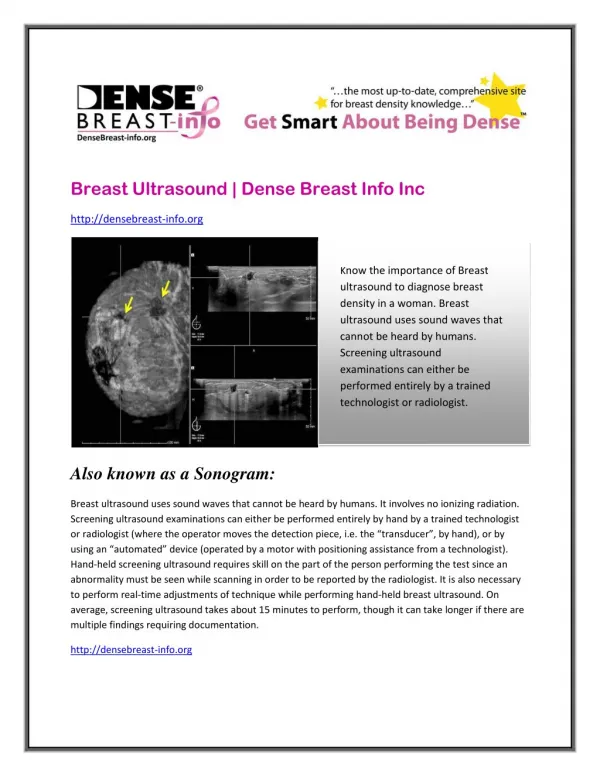
9. Breast cancer screening • Decrease sensitivity of dense breast on screening mammogram • US can be used in addition to find underlying mass
Ultrasound technique • lighting • patient positioning: support elbow, flat, supine • ergonomics • probe: linear array 7-13 MHz • scanning: radial/antiradial • clock face with distance from nipple • only caliper things that are REAL • correct depth (skin to pectoral fascia) and correct focal zone (up to 2 is OK) • dynamic range: some settings can make a cystic lesion look solid and vice versa
Ultrasound interpretation • Breast Composition • Mass • Calcifications • Associated features
Breast composition • composition of breast tissue • Glandular tissue (soft tissue density) • Connective tissue (soft tissue density) • Fat (fat density) Young women : glandular tissue > fat Older women : fat > glandular tissue More glandular tissue dense breast on mammogram
Breast Composition: • Homogeneous echotexture - fat • Homogeneous echotexture - fibroglandular • Heterogeneous echotexture
Mass • Shape • Margin • Orientation • Echo pattern • Posterior features
Breast mass Shape
Breast mass Margin
Orientation: • unique to US-imaging, and defined as parallel (benign) or not parallel (suspicious finding) to the skin.
Echo pattern • Anechoic • Hypoechoic, • Complex cystic • Solid:isoechoic, hyperechoic, heterogeneous.
Posterior features: • Enhancement • Shadowing.
Calcifications: • On US poorly characterized compared with mammography, but can be recognized as echogenic foci, particularly when in a mass.
Associated features: • Architectural distortion • Duct changes • Skin changes • Edema • Vascularity • Elasticity assessment
Findings suggestive of malignancy Spiculation or thick echogenic halo Angular margin Microlobulation Shape taller than wide Duct extension or branch pattern Acoustic shadowing Microcalcifications Hypoechogenicity
Findings suggestive of benign • No suggestive findings of malignancy • Hyperechogenicity (compare to fat) • Wider than tall • Well circumscribed, less than 3 lobulations • Thin echogenic pseudocapsule
Ultrasound Features Suggestive of Benign and Malignant Nodules.
Special cases cases with a unique diagnosis or pathognomonic ultrasound appearance: • Simple cyst • Complicated cyst • Clustered microcysts • Mass in or on skin • Foreign body including implants • Lympnodes- intramammary • Lymph nodes- axillary • Vascular abnormalities • Postsurgical fluid collection • Fat necrosis
BreastImagingReportingAndDataSystem(BI-RADS) Mammography • Category 0 ...incomplete assessment additional imaging is needed • Category I ...negative routine follow up (1 year) • Category II … benign finding routine follow up (1 year) • Category III … probably benign finding follow up 6 months • Category IV … suspicious abnormality biopsy • Category V… highly suggestive of malignancy biopsy • Category VI …known biopsy proven malignancy appropriate treatment Help clinicians for management