Download

1 / 23
280 likes | 584 Vues
The role of ultrasound in breast imaging. Dr Francien Malan Drs Van Wageningen & Vennote 31 October 2007. How does ultrasound work?. High frequency sound wave Crystal probe serves as both transmitter and detector of sound waves Different tissue types

E N D
The role of ultrasound in breast imaging Dr Francien Malan Drs Van Wageningen & Vennote 31 October 2007
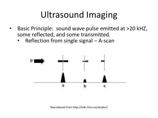
How does ultrasound work? High frequency sound wave Crystal probe serves as both transmitter and detector of sound waves Different tissue types Signal coming back translated into real time black and white picture by computer software
Uses of ultrasound in breast imaging • Palpable masses • Mammographically detected masses • Dense breasts • Young patients • Pregnant/ lactating woman • Breast implants • Guided aspiration/ biopsy/ localisation
Palpable abnormality • Ultrasound especially useful if mammogram shows no obvious abnormality • +/- mammo shows abnormality • Young patients • Benefits of ultrasound
Simple cyst Typical fibroadenoma
Dense/whitebreasts Fatty/ dark breasts
Dense breasts means a relatively large percentage of fibroglandular tissue and little fat • 50% of patients <30yrs • 1/3 of patients > 50yrs • Can’t “see through” • Ultrasound useful!!!
Young patients (<30/ <35yrs) • Should be first investigation; mammogram only if ultrasound equivocal • Palpable lesions in young woman most commonly cysts or fibroadenomas
Most common problem in lactating woman is mastitis +/ breast abcesses • + US guided drainage of abcess
Indications the same as for women without implants • Also for evaluation of implant complications such as rupture
Ultrasound guided cyst aspiration/ biopsy • Aspiration of cysts are done when cyst has atypical features, pain relief, relief of anxiety, cosmetic reasons • Biopsy done when after clinical evaluation/ mammography and ultrasound the nature of lesion is still uncertain
What happens? • Outpatient • Sterilised, anaethetised • Needle is guided into cyst under direct ultrasound vision • Cells obtained to path lab for evaluation
Ultrasound guided localisation • Localisation is done prior to surgical resection of lesion to guide surgeon to the lesion, can be done with u/s or mammogram • Inpatient, fasting, sterile conditions, local anaesthetic, localisation needle guided into lesion, wire strapped to arm, patient goes to theatre.
Why ultrasound for localisation? • Lesion visible on ultrasound, not clinically palpable; may or may not be visible on mammogram • Benefits of real time guidance of wire into lesion; 3D perspective; relatively quick;
Why mammogram for localisation? • Microcalcifications • Small lesion deeply seated in large breasts • Area of suspicion on mammogram not visible on ultrasound
Complications • Unsuccessful • Hematoma • Minor discomfort • Infection
Limitations of breast ultrasound • Many cancers are not visible on ultrasound • Microcalcifications • Inderteminate > biopsy
CANNOT REPLACE REGULAR SELF EXAMINATION AND MAMMOGRAPHY AS PRIMARY SCREENING TOOL FOR BREAST CANCER!!!!