Download

1 / 17
1.84k likes | 9.07k Vues
Basic Principles of Ultrasound Imaging. Introduction Ultrasound imaging have different meaning to to different categories of people based on profession or vocation In the Heath Sector- Clinicians, Patients

E N D
Basic Principles of Ultrasound Imaging Introduction Ultrasound imaging have different meaning to to different categories of people based on profession or vocation In the Heath Sector- Clinicians, Patients In Commerce- Business & Industry Definition: Ultrasound Imaging (Scanning) • Commonly referred to as “Scanning,,” is the act of acquiring and interpreting images using sound waves • Medically : Involves the use of high frequency ( higher than human hearing) sound waves to produce images of internal organs of the body. • Technically, it is a noninvasive medical procedure that allow internal structures of the body and hemodynamics of blood flow to be accessed or viewed as real-time images on a screen. • Ultrasound transducers send out high frequency sound waves into the body tissues and receive reflected echoes , that are processed and displayed as images. • For diagnostic purposes Ultrasound Doppler frequency range is 2.0 -10.5 MHz . The frequency range for scanners for industrial or commercial use is different • Normal hearing sound wave frequency for adults is 2.5- 3.5 MHz
Basic Facts: One major fact about Ultrasound Imaging(Scanning) that distinguishes it from other Non Invasive Diagnostic technology tools in the health industryis that... • “ The accuracy of the noninvasive examination is almost exclusively dependent on the skill and the experience of the operator.’’ Therefore, if the Operator misses the disease during the process of scan, No matter how knowledgeable the interpreter (Reader) is, it is unlikely that it will not be detected unless... Other diagnostic tests were ordered • This places a unique responsibility on the operator to strive and gain the knowledge and competency required, as patient mistreatment / misdiagnosis due to professional incompetence and performance is more dangerous than the disease itself..
Overview of Presentation Part 1 Review Basic Ultrasound Principles • Definitions and Terminologies • Ultrasound Physics • The Transducer ( Probe ) • Review Ultrasound Modalities in Cardiovascular Testing Part 2 • I Demonstrations • Exercises • Probe Handing , Orientation and Scanning techniques • Scanning to identify body organs
Objectives • Update awareness of various Ultrasound tests available • Enhance professional skills acquisition by Physicians to enable proper evaluation of scanning reports or images given to patients. • Enhance appropriate use of ultrasound systems in patient management • Improve overall quality of patient care delivery through accurate diagnosis
Descriptive Definitions Terminologies A • Cephalad / caudal (toward the head/toward the tail): used interchangeably with superior and inferior. • Superior / inferior (above/below): for location of a structure along the long axis of the body. • Dorsal / ventral (backside / belly side): ventral always refers to the belly side . • Proximal / distal(nearer the trunk or attached end. • Superficial / deep (toward or at the body surf ace / away from the body surface or more internal). • Anterior / posterior (front / back): anterior structures are those that are most forward—the face, chest, and abdomen. • Medial/lateral (toward the midline / away from the midline or median plane):
Descriptive Definitions Terminologies Body Planes Imaginary surface or line called a plane that lie at right angles to one another. • Sagittal plane: divides the body into equal parts, right down the median plane of the body, it is called a median, or mid sagittal plane. • Frontal (coronal) plane: divides the body (or an organ) into anterior and posterior parts. • Transverse plane: divides the body into superior and inferior parts. The terms above assume the person is in the anatomical position
Descriptive DefinitionTerminologies Sonography • ANECHOIC - Being echo-free or without echoes (e.g., a fluid-filled cyst). • ECHOGENICITY- Echogenic: the ability to create an ultrasound echo . • ECHOLUCENT- same as above. • HETEROGENEOUS - mixed echoic pattern within plaque- areas of sonolucence and echogenicity. • HOMOGENEOUS - uniform plaque texture. • HYPERECHOIC - Producing echoes of higher amplitude than normal for the surrounding medium. • HYPOECHOIC - Producing echoes of lower amplitude than normal for the surrounding medium. • ISOECHOIC- Areas which have similar echogenicity to each other. An isoechoic "property" makes it more difficult to see the desired tissue structure. • SONOLUCENT- Allowing passage of ultrasonic waves without echoes
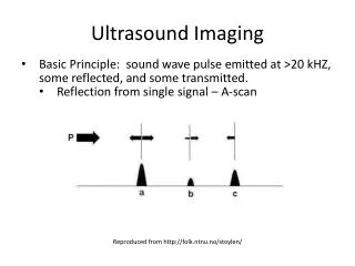
Ultrasound PhysicsEcho Doppler Principle • Mechanism of Sound Wave Generation: • Ultrasound transducers have elements made of Piezoelectric • crystals. • Major Kinds of Transducers Generated Doppler Waves • Continuous Wave (CW) Doppler and Pulse Wave(PW) Doppler • When stimulated with electricity , the crystals oscillate to produce a • high frequency sound wave signal. • Transducers send sound out and receive returning echoes from • moving reflectors in the body ( Blood and tissues ). • The wave frequency difference between the transmitted waves and • the reflected waves due to movement is called • Doppler Shift frequency • If the returning wave frequency is lower than the transmitted frequency, the Doppler shift is considered a NEGATIVE (-) • If the returning frequency is higher, the Doppler shift is POSITIVE (+)) Dopplers and Pulse Wave (PD)
Doppler Shift Echo Doppler transmission is the principle mechanism use in Ultrasound systems to detect and measure blood flow dynamics . • Doppler shift frequency is expressed as a positive or negative value, depending on the direction of flow relative to the Doppler beam direction
Ultrasound physicsThe Doppler Equation • The Doppler Equation is used to calculate blood flow velocity if the speed of sound in tissue is known as well as the angle between blood flow and the ultrasound beam. The Equation follows V= C (±Δf ) Where 2 foCosѲ V= Blood flow velocity (meters/s) C = Speed of sound in tissue; approx 1540mm/s Δf =Doppler frequency Shift (Hz) fo= transmitted frequency CosѲ= Cosine function of angle between ultrasound beam and the blood flow vector
Doppler Wave formParameters Pulse Wave Doppler(PW) • have single crystal to send &receive transmission • Range Gate (TGC) (Sample Volume) at any specific depth. - Sets Time interval in PW Mode . Amplified contrast of reflected sound waves to form images • Pulse Repetition Interval(PRF) is number of pulse echo cycles per second • PRF is expressed in Hertz; • 11/2 PRF = Nyquist Limit • Aliasing occurs when Doppler shift frequency exceeds 11/2 of PRF. • To reduce Aliasing; increase PRF; Decrease frequency; Decrease sample volume depth volume • Wall Filters (Hz) are set to detect or eliminate low flow As PRF increases; Wall filters increases
The Scanning Techniques Protocols “Standard" Protocol remain unchanged if proven • To be effective, • Cost efficient and • accurate, • Have been no better replacements. Protocols change as • Better methods evolve • Technological advances • New technologies mandate new test methods
Applications Angleof Insonation Range 45 ⁰ To 60⁰
Applications of Ultrasound Imaging in Cardiovascular Testing Stroke Screening Carotid plaque estimation Carotid Artery Duplex Examinationn CVA stenosis evaluation Peripheral Arterial Disease (PAD) Imaging Segmental pressure scan stdiesforr localization Plethysmography-Skin per Fusion evaluation of blood flow with IFR photosensors Venous Duplex Scan for Thrombosis Duplex studies for blood clot in vessels Trans Cranial Imaging Meningeal arterial flow studies Abdominal Aorta Imaging for Aneurysms Abdominal scan for aneurysms Renal Vascular Studies Renal vascular flow studies
In Conclusion • Ultrasound scanning has become the most affordable diagnostic tool in our hospitals and clinics today. • There is need for professional improvement on theoverall quality of patient care delivery through accurate diagnosis and knowledge based evaluation of patient reports from Scanning Facilitiesby physicians . Thank You.
REFERENCES • Johnston KW, Rutherford RB, Tilson MD, et al. Suggested standards for reporting on arterial aneurysms. J Vasc Surg. 1991;13:452-458 • McConathy WJ, Alaupovic P, Woolcock N, et al. Lipids and apolipoprotein profiles in men with aneurysmal and stenosingaorto-iliac atherosclerosis. Fur J Vasc Surg. 1989;3:511-514 • Emedicine- http://www.emedicinehealth.com • Meyers P, Owens C, Renal Doppler. Integrated Ultrasound Reference Guide, SDMS Educational Foundation, Dallas TX 1996 • Kohler TR, Zeirler RE, Martin RL et al. Nonivasive Diagnosis Of Renal Artery Stenosis By Ultrasonic Duplex Scanning. J VascSurg 1986;4;450-6. • Taylor DC, Kettler MD, Moneta GL, et.al, Duplex Ultrasound scaniing in the diagnosis of renal artery stenosis: A prospective evaluation. J VascSurg 1988;7:363-9 • Hansen KJ,Tribble RW, Reavis SW, et. al., Renal duplex sonography: Evaluation of clinical utility. J VascSurg 1990;12:227-36 • Martin RL, Nanra RS, Bray AE, et al., Renal Hilar Doppler analysis in the Detection of Renal Artery Stenosis. J VascTechnol 15(4):173-180 1991 • Stavros AT, Harchfield D,. Renal Doppler, renal artery stenosis, and renovascular hypertension: direct and indirect sonographic abnormalities in patients with renal artery stenosis.Ultrasound Quarterly, Vol.12,No.4, pp. 217-263, 1994 Raven Press Ltd, New York • Kotval PS. Doppler Waveform parvus and tardus: a sign of proximal flow obstruction. J Ultrasound Med 1989;8:435-40. • Rene CE, Oliva VL, Bui BT et. al., Renal Artery Stenosis: Evaluation of Doppler US after Inhibition of Angiotensen - converting enzyme with Captopril. Radiology 1995; 196:675-679.