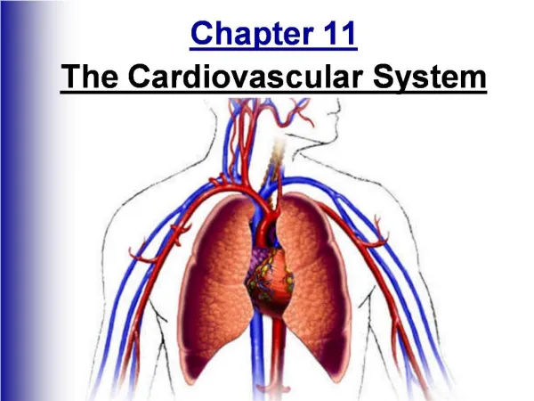
Ch 11 Cardiovascular System
Ch 11 Cardiovascular System. Cardiovascular System: The Heart 1. State the function of the cardiovascular system and its components. 2. Describe the location of the heart in the body and identify its major anatomical areas on an appropriate model or diagram.

Ch 11 Cardiovascular System
E N D
Presentation Transcript
Ch 11 Cardiovascular System Cardiovascular System: The Heart 1. State the function of the cardiovascular system and its components. 2. Describe the location of the heart in the body and identify its major anatomical areas on an appropriate model or diagram. 3. Trace the pathway of blood through the heart. 4. Compare and contrast the pulmonary and systemic circuits. 5. Explain the operation of the heart valves. 6. Name the functional blood supply of the heart. 7. Name the elements of the intrinsic conduction system of the heart and describe the pathway of impulses through this system. (SA node, AV node…) 8. Define systole, diastole, stroke volume, and cardiac cycle. 9. Define heart sounds and murmur. Cardiovascular System: Blood Vessels 11. Compare and contrast the structure and function of arteries, veins, and capillaries. 12. Identify the body's major arteries and veins. 13. Define blood pressureand list factors affecting and/or determining blood pressure. 15. Define hypertension, atherosclerosis and varicose veins & describe possible health consequences of these conditions. 16. Describe the exchanges that occur across capillary walls. 17. Name the fetal vascular modifications, or "fetal shunts," and describe their function before birth. 18. Explain how regular exercise and a diet low in fats and cholesterol may help maintain cardiovascular health.
Just to get you thinking about what you already know about the cardiovascular system Write a paragraph that explains what you think the function of the cardiovascular system is, it’s parts, and anything you know about the structure of the parts. (Think about things you’ve learned this year that you can apply to this and include them.) Draw a picture of the heart and label it. Here’s a little something to get you in the mood:
Functions & Parts? Your heart pumps 6 L of blood from your heart to you blood vessels & back over 1000 times / day! • Open heart surgery video: http://www.youtube.com/watch?v=Zxqj1BcBpIg Explanation of bypass surgery: http://www.youtube.com/watch?v=WM_tcf5Ogy0 Video of off-pump bypass surgery: http://www.youtube.com/watch?v=MGzyhuCs43o
Learning the part of the heart • Practice: • External: http://wps.aw.com/bc_marieb_ehap_8/25/6528/1671273.cw/index.html • Internal: http://wps.aw.com/bc_marieb_ehap_8/25/6528/1671273.cw/index.html
The Cardiovascular System – Goal 1 • Consists of ______& blood ________ • Function of heart is to______________ • Function of blood vessels is to ________blood around body • The functions of the cardiovascular system • To deliver_____,_______, hormones to tissues • To remove ______& other ______from tissues
Location – Goal 2 Midsternal line 2nd rib Sternum Diaphragm Point of maximal intensity (PMI) (a) Figure 11.1a
Goal 2 Superior vena cava Aorta Parietal pleura (cut) Pulmonary trunk Left lung Pericardium (cut) Apex of heart Diaphragm (c) Figure 11.1c
The Heart – Goal 2 • Location • Thorax between the _____in the inferior___________ • Orientation • Pointed ______directed toward left______ • Base points toward right_______ • About the size of your ______
The Heart: Coverings – Goal 2 • Pericardium—a double-walled ____ • Video showing pericardium getting cut during surgery • http://www.youtube.com/watch?v=hchNmUsx7hQ
The Heart: Chambers – Goal 2 • Right and left side act as separate _____ • ___ chambers • 2 ________ • Receiving chambers • Right atrium • Left atrium • 2 _________ • Discharging chambers • Right ventricle • Left ventricle
The Heart: Septa Goal 2 • Interventricular septum • Separates the two _________ • Interatrialseptum • Separates the two______ Left ventricle Right ventricle Muscular interventricular septum Figure 11.5
The Heart: Associated Great Vessels – Goal 2 • Arteries • Aorta • Leaves_____ __________ • Pulmonary arteries • Leave______ __________ • Veins • Superior and inferior venaecavae • Enter ______ _________ • Pulmonary veins (four) • Enter ________ ________
Superior vena cava Aorta Left pulmonary artery Right pulmonary artery Left atrium Right atrium Left pulmonary veins Right pulmonary veins Pulmonary semilunar valve Left atrioventricular valve (bicuspid valve) Fossa ovalis Aortic semilunar valve Right atrioventricular valve (tricuspid valve) Left ventricle Right ventricle Chordae tendineae Interventricular septum Inferior vena cava Myocardium Visceral pericardium (b) Frontal section showing interior chambers and valves. Figure 11.3b
Links to practice sites: • http://wps.aw.com/bc_marieb_ehap_8/25/6528/1671273.cw/index.html
The Heart’s Role in Blood Circulation – Goal 3 & 4 • Systemic circulation • Blood flows from the _____ side of the heart through the ______ tissues and back to the _____ side of the heart • Pulmonary circulation • Blood flows from the _____ side of the heart to the ______ and back to the _____ side of the heart.
Blood Flow Through the Heart – Goal 3 • ________and ________ venaecavae dump blood into the _______ ________ • From right atrium, through the ________ valve, blood travels to the ________ ____________ • From the right ventricle, blood leaves the heart as it passes through the ___________ semilunar valve into the ___________ trunk • Pulmonary trunk splits into right and left pulmonary ________ that carry blood to the ________ Continued on next page…
Blood Flow Through the Heart – Goal 3 • _________ is picked up and _______ ________ is dropped off by blood in the lungs. • Oxygen-rich blood returns to the heart through the four _________ _______. • Blood enters the left ______ and travels through the _________ valve into the left _____________ • From the left ventricle, blood leaves the heart via the _______ semilunar valve and aorta • http://www.youtube.com/watch?v=rguztY8aqpk
The Heart’s Role in Blood Circulation – Goal 3 & 4 • In both the pulmonary and systemic circulations, the exchange of _________, nutrients, and _________ products occurs in the capillaries that join arterioles to venules
The Heart: Valves – Goal 5 • Allow blood to flow in only one direction to prevent ___________ • Four valves in heart • Atrioventricular (AV) valves—between _____and__________ • ___________ valve (right side of heart) • __________ (mitral) valve (left side of heart) • Semilunar valves—between ventricle and ______ • Pulmonary semilunar valve • Aortic semilunar valve
Superior vena cava Aorta Left pulmonary artery Right pulmonary artery Left atrium Right atrium Left pulmonary veins Right pulmonary veins Pulmonary semilunar valve Left atrioventricular valve (bicuspid valve) Fossa ovalis Aortic semilunar valve Right atrioventricular valve (tricuspid valve) Left ventricle Right ventricle Chordae tendineae Interventricular septum Inferior vena cava Myocardium Visceral pericardium (b) Frontal section showing interior chambers and valves. Figure 11.3b
The Heart: Valves – Goal 5 • AV valves • __________ in place by chordaetendineae (“heart strings”) • ________ during heart relaxation and ________ during ventricular contraction • Semilunar valves • _________ during heart relaxation but _________ during ventricular contraction • Notice these valves operate __________ of one another to force a _____-way path of blood through the heart
Cardiac Circulation - Goal 6 • Blood in the 4 chambers of the heart does not nourish the myocardium… so what does?
Cardiac Circulation - Goal 6 • Cardiac circulation supplies the myocardium with blood. • The heart has its own nourishing circulatory system consisting of • Coronary __________—branch from the aorta to supply the heart muscle with _______________ blood • Cardiac _________—drain the myocardium of blood • Coronary sinus—a large vein on the posterior of the heart, receives blood from cardiac veins • Blood empties into the right atrium via the coronary sinus
Homeostatic Imbalance – Goal Bonus • Angina pectoris – crushing pain from __________ blood flow to myocardium. May be experienced when heart beats at a rapid pace (coronary flow occurs when heart is relaxed but if heart is beating rapidly there is little relaxation time) This is a warning that should not be ignored. • Myocardial infarction (aka “heart attack” or “coronary”) – ________ of oxygen deprived heart cells.
Women are more likely than men to have the “other” signs of a heart attack. • Call 911 – get to the hospital as quickly as possible.
The Heart: Conduction System – Goal 7 • Intrinsic conduction system (nodal system) • Heart muscle cells contract, without______ ________, in a regular, continuous way (Indiana Jones video below) • http://www.youtube.com/watch?v=KBIdcUxdgo0 • Special tissue sets the pace • Sinoatrial node = SA node (“_________”), is in the right _______ • Atrioventricular node = AV node, is at the junction of the atria and _____________
The Heart: Conduction System – Goal 7 Superior vena cava Sinoatrial (SA) node (pacemaker) Left atrium Atrioventricular (AV) node Atrioventricular (AV) bundle (bundle of His) Right atrium Bundle branches Purkinje fibers Interventricular septum Purkinje fibers Figure 11.7
Homeostatic Imbalance – Goal Bonus • Heart block—damaged AV node releases ventricles from control of the SA node; result is in a slower heart rate as ventricles contract at their own rate • Ischemia—lack of adequate _______ supply to heart muscle • Fibrillation—a ______, uncoordinated shuddering of the heart muscle • Video that shows various types of arrhythmias • http://www.bupa-intl.com/health/factsheets/H/Heart-block#tabContainer
Homeostatic Imbalances – Goal Bonus • Homeostatic imbalance (continued) • Tachycardia—______ heart rate over 100 beats per minute • Bradycardia—______ heart rate less than 60 beats per minutes
The Heart: Cardiac Cycle & Heart Sounds – Goal 8 • Atria contract _____________ • Atria relax, then ___________contract • Systole = ventricular____________ • Diastole = ventricular _____________
The Heart: Cardiac Cycle & Heart Sounds Goal 8 & 9 Cardiac cycle—events of one complete heart beat Heart Sounds: • ____ - Atrioventricular valves close causes first heart sound • ____ - Second heart sound is heard as semilunar valves close • Murmur – _______ heart sounds due to leaky valves or other issues.
The Heart: Cardiac Output – Goal 8 (SV) • Cardiac output (CO) • Amount of blood pumped by each side (ventricle) of the heart in one minute • Determined by SV x HR • Stroke volume (SV) • _________ of blood pumped by each ventricle in one contraction (each heartbeat) • About 70 mL of blood is pumped out of the left ventricle with each heartbeat • Heart rate (HR) • Typically 75 beats per minute
The Heart: Regulation of Heart Rate • Increased heart rate • Crisis • Low blood pressure • Hormones • Epinephrine • Thyroxine • Exercise • Decreased blood volume • Decreased heart rate • Parasympathetic nervous system • High blood pressure or blood volume • Decreased venous return
Blood Vessels: The Vascular System – Goal 11 • Transport blood to the tissues and back to heart. • Carry blood _______ from the heart • Arteries • Arterioles • Exchanges between tissues and blood • ___________ – one cell thick • Return blood _______ the heart • Venules • Veins - valves
Artery Vein (a) Figure 11.10a
Venous Aids for the Return of Blood to the Heart – Goal 11 Valve (open) • To assist in the movement of blood back to the heart: • Larger veins have _______ to prevent backflow • __________ muscle “milks” blood in veins toward the heart Contracted skeletal muscle Valve (closed) Vein Direction of blood flow Figure 11.11
Vascular shunt Precapillary sphincters True capillaries Terminal arteriole Postcapillary venule Sphincters open; blood flows through true capillaries. Figure 11.12a
Anterior Cerebral arterial circle (circle of Willis) Frontal lobe Optic chiasma • Anterior communicating artery Middle cerebral artery • Anterior cerebral artery Internal carotid artery • Posterior communicating artery Mammillary body • Posterior cerebral artery Temporal lobe Basilar artery Vertebral artery Pons Occipital lobe Cerebellum Posterior (a) Figure 11.15a
Ductus arteriosus Superior vena cava Pulmonary artery Pulmonary veins Foramen ovale Inferior vena cava Hepatic vein Ductus venosus Inferior vena cava Hepatic portal vein Umbilical vein Fetal umbilicus Aorta Common iliac artery Umbilical cord External iliac artery Internal iliac artery Umbilical arteries Urinary bladder KEY: High oxygenation Moderate oxygenation Low oxygenation Very low oxygenation Placenta Figure 11.16
Fetal Circulation • Fetus receives exchanges of gases, nutrients, and wastes through the placenta • Umbilical cord contains three vessels • Umbilical vein—carries blood rich in nutrients and oxygen to the fetus • Umbilical arteries (2)—carry carbon dioxide and debris-laden blood from fetus to placenta
Fetal Circulation – Goal 17 Blood flow bypasses the lungs • Foramen ovale - ____________________ • Ductusarteriosus connects the aorta and pulmonary trunk (becomes ligamentumarteriosum at birth)
Left common carotid artery Brachiocephalic trunk Left subclavian artery Superior vena cava Aortic arch Right pulmonary artery Ligamentum arteriosum Ascending aorta Left pulmonary artery Pulmonary trunk Left pulmonary veins Left atrium Right pulmonary veins Auricle of left atrium Right atrium Circumflex artery Right coronary artery in coronary sulcus (right atrioventricular groove) Left coronary artery in coronary sulcus (left atrioventricular groove) Anterior cardiac vein Left ventricle Right ventricle Great cardiac vein Marginal artery Anterior interventricular artery (in anterior interventricular sulcus) Small cardiac vein Inferior vena cava Apex (a) Figure 11.3a
Superior vena cava Aorta Left pulmonary artery Right pulmonary artery Left atrium Right atrium Left pulmonary veins Right pulmonary veins Pulmonary semilunar valve Left atrioventricular valve (bicuspid valve) Fossa ovalis Aortic semilunar valve Right atrioventricular valve (tricuspid valve) Left ventricle Right ventricle Chordae tendineae Interventricular septum Inferior vena cava Myocardium Visceral pericardium (b) Frontal section showing interior chambers and valves. Figure 11.3b