Photon Tissue Interactions
Photon Tissue Interactions . The Electromagnetic Spectrum (EMS) . light. Wavelength refers to the distance between two consecutive wave crests Frequency refers to the number of cycles per second (cps); its unit of measure is the hertz (Hz), which is equal to 1 cps.

Photon Tissue Interactions
E N D
Presentation Transcript
Wavelength refers to the distance between two consecutive wave crests • Frequency refers to the number of cycles per second (cps); its unit of measure is the hertz (Hz), which is equal to 1 cps. • Frequency and wavelength are closely associated with the relative energy of electromagnetic radiations. More energetic radiations have shorter wavelengths and higher frequency. The relationship among frequency, wavelength, and energy is illustrated in the electromagnetic spectrum.
IMAGE CREATION • ATOMS • INTERACTION WITH “MATTER” • ATOMIC NUMBER
Interaction in The body begin at the atomic level Atoms Molecules Cells Tissues Organ structures
No interaction; X-ray passes completely through tissue and into the image recording device. Complete absorption; X-ray energy is completely absorbed by the tissue. No imaging information results. Partial absorption with scatter; Scattering involves a partial transfer of energy to tissue, with the resulting scattered X-ray having less energy and a different trajectory. Scattered radiation tends to degrade image quality and is the primary source of radiation exposure to operator and staff. Interactions of X-rays with matter
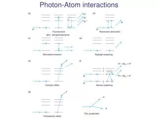
Patient Interactions • **Photoelectric** • Classic Coherent Scatter • **Compton Scattering** • Pair Production • Photodisintegration
Coherent Scattering • Also called: Classical scattering or Thompson scattering • Occurs with energies below 10 keV • Incident photon interacts with an atom of matter, causing it to become excited. Immediately the atom releases this excess energy and a new scattered photon. • The wavelength is equal to the incident x-ray or equal energy. • The only difference is the direction of travel
Classical (Coherent) Scattering • Excitation of the total complement of atomic electrons occurs as a result of interaction with the incident photon • No ionization takes place • Electrons in shells “vibrate” • Small heat is released • The photon is scattered in different directions • No loss of E
Compton Effect or Compton Scattering • Occurs throughout the diagnostic imaging range • The incident x-ray interacts with the outer electron shell on an atom of matter, removing it. • It not only causes ionization but scatters the incident x-ray causing a reductions in energy and the change of direction.
Compton scatter • A fairly high energy (high kVp) x-ray photon ejects an outer shell electron. • Though the x-ray photon is deflected with somewhat reduced energy (modified scatter), it retains most of its original energy and exits the body as an energetic scattered photon. • A Compton e- is also released • Since the scattered photon exits the body, it does not pose a radiation hazard to the patient. • It can, however, contribute to film fog and pose a radiation hazard to personnel (as in fluoroscopic procedures).
Compton scatter • Both the scattered x-ray and the Compton electron have enough energy to cause more ionization before loosing all their energy • In the end the scattered photon is absorbed photoelectrically • Compton is just as likely to occur with soft tissue as bone. • Compton is significant • Scattered photons provides no useful diagnostic information
Compton Effect • The Compton electron looses all of its kinetic energy by ionization and excitation and drops into a vacancy in an electron shell previously created by some other ionizing event • The probability of Compton effect increasesas photon energy increases, however the atomic number does not affect the chances of the Compton effect
Compton Effect • Scattered radiation produces a uniform optical density on the radiograph that reduces image contrast • Scattered radiation from Compton contributes to the majority of technologists exposure, especially during fluoroscopy
During Fluoroscopy – the patient is the largest scattering object
Radiation Protection during Fluoro • The least exposure is 90 degrees to the patient • The most exposure is at the head or feet of the patient table
Photoelectric Effect or Absorption • Inner-shell ionization • The photon is not scattered it is totally absorbed • The e- removed from the atom of matter is called a photoelectron, with an energy level equal to the difference between the incident photon and the e- binding energy.
PHOTOELECTRIC ABSORBTION IN THE PATIENT (CASCADE OF ELECTRONS)
Photoelectric effect • A relatively low energy (low kVp) x-ray photon uses all its energy (true absorption) to eject an inner shell electron, • leaving an orbital vacancy. • An electron from the shell above drops down to fill the vacancy and, in doing so, gives up energy in the form of a characteristic ray. • The photoelectric effect is more likely to occur in tissues with high atomic numbers (eg, bone, positive contrast media) • and contributes significantly to patient dose, • as all the photon energy is absorbed by the patient (and for the latter reason, is responsible for the production of short-scale contrast).
Electron transitions • Are accompanied by the emission of more x-rays – secondary radiation • Secondary radiation behaves much like scatter radiation • Secondary contributes nothing to the image • The probability that any given photon will undergo a photoelectric interaction is dependent on the photon energy and the atomic number of the atom
PHOTOELECTRIC ABSORBTION IS WHAT GIVES US THE CONTRAST ON THE FILM
Important X-ray Interactions • Of the five interactions only two are important to radiology • Photoelectric effect or photoelectric absorption • Compton scatter • Which two tube interactions are important • Draw them
Compton scatter • Contributes to no useful information • Is independent of the atomic number of tissue. The probability of Compton is the same for bone atoms and for soft tissue atoms • The probability for Compton is more dependent on kVp or x-ray energy
Compton Scatter • Results in image fog by optical densities not representing diagnostic information • Photon are Photons IR is does not know the difference
Photoelectric Absorption • Provides information to the IR because photons do not reach the IR • This represents anatomic structures with high x-ray absorption characteristics; radiopaque structures; tissue with high atomic number; or tissue with high mass density
Attenuation – The total reduction in the # of photons remaining in an x-ray beam after penetration through tissue • Absorption = x-ray disappears (Photoelectric, Pair production & Photodisintegration) • Scattering = partially absorbed, x-ray emerges from the interaction traveling in a different direction (sometimes with less energy) • Absorption + Scattering = Attenuation
IMAGE FORMATION • DIFFERENTIAL ABSORPTION = the difference between those x-rays absorbed and those transmitted to the IR • Compton scatter (no useful information) • Photoelectric absorption (produces the light areas on the image) • Transmitted x-rays (produces the grey/dark areas on the image)
The probability of radiation interaction is a function of tissue electron density/ atomic number, tissue thickness/density, and x-ray energy (kVp). • Dense material like bone and contrast dye attenuates more X-rays from the beam than less dense material (muscle, fat, air). • The differential rate of attenuation provides the contrast necessary to form an image.
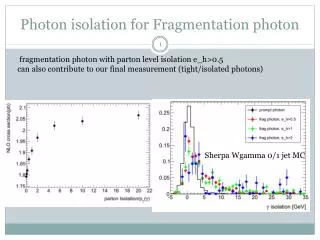
Differential Absorption • Increases as the kVp is reduced • Approximately 1% of photons that interact with the patient (primary beam) reach the IR. Of that 1% approximately 0.5% interact to form the image
Compton vs. Photoelectric • Below 60 kVp Photoelectric absorption is predominant above 60 kVp Compton scatter begins to increase. • Dependent on the tissue attenuation properties
Differential absorption factors • High atomic number = larger atoms • Mass Density = how tightly the atoms of tissue are packed • Z # for air and soft tissue are about the same the OD changes are due to mass density difference • Table 12–3 & 12-5