Download
1 / 56
560 likes | 653 Vues
Explore the anatomy, physiology, and functions of the nervous system, including sensory, integrative, and motor functions, along with details on neurons, synapses, and divisions of the nervous system.

E N D
NERVOUS SYSTEM INTRODUCTION BIO 137 Anatomy & Physiology I
Nervous System • One of body’s 2 main control systems • Detects a stimulus inside or outside of body & coordinates a response • Acts by electrical signals, called nerve impulses • Nervous tissue is excitable • Controls most of body’s activities to help maintain homeostasis
Organs of the Nervous System • Brain • Spinal Cord • Nerves • Cranial • Spinal • Peripheral
Functions of the Nervous System • 3 main functions • Sensory function • Integrative function • Motor function
Sensory Function of the Nervous System • Sensory receptorsare located at the ends of peripheral nerves • Sensory receptors detect a change outside or inside the body • Monitor internal, external and environmental factors • Light and sound, temperature, oxygen concentration • Information from sensory receptors is converted into a nerve impulse, which is transmitted from peripheral nerves to the CNS
Integrative Function of the Nervous System • The nervous system processes sensory information by analyzing it and making decisions for an appropriate response • ‘Sensory information is integrated‘
Motor Function of the Nervous System • Conscious and subconscious decisions are made and acted upon • Response is carried out by effectorsthrough cranial and spinal nerves • Muscles and/or glands
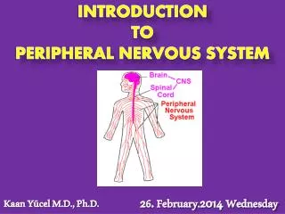
Divisions of Nervous System • Central Nervous System, CNS • Brain • Spinal cord • Peripheral Nervous System, PNS • Peripheral nerves • Cranial nerves • Spinal nerves • PNS connects CNS to body parts
Peripheral Nervous System Divisions • Sensory Division • Sensory receptors in the PNS pick up sensory information from the body and deliver it to the CNS as a nerve impulse • Motor Division • Neurons carry information from the CNS to effectors
PNS: Motor Division • SomaticDivision • Oversees voluntary activities • Skeletal muscle contraction • Autonomic Division • Controls visceral activities • Involuntary activities • Including: Heart rate, blood pressure, breathing, body temperature
PNS:Motor:Autonomic • Sympathetic • Active under stressful situations • ‘Fight or Flight’ • Parasympathetic • Active under normal, restful conditions • Returns body to homeostatic levels following a stressful experience
Divisions of the Nervous System • Ganglia are small masses of neuronal cell bodies located outside the brain and spinal cord, usually closely associated with cranial and spinal nerves. • There are ganglia which are somatic, autonomic, and enteric (that is, they contain those types of neurons.)
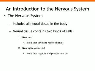
Histology of Nervous System • 2 main types of cells • Neurons • A single nerve cell • Neuroglia • Support and nutrition of brain cells • ‘neuron helpers’ • Found in both CNS and PNS
Neuronal Structure • Dendrite(s)– receive information from within or outside the body • Cell body– contains nucleus, organelles • Axon hillock - Axon arises from here on the cell body • Axon– transmits received information as a nerve impulse • A neuron may have many dendrites but only 1 axon • Axon may have branches towards its end called collaterals • Ends of each axon collateral are called axon terminals which end at a synaptic knob • Vesicles of neurotransmitter located here • Synapse exists between end of an axon and what it innervates
Flow of Information in a Neuron • Dendrite(s) • Cell Body • Axon hillock • Axon • Collaterals • Axon Terminals • Synaptic knob
Axons can terminate at three locations 1. Muscle 2. Gland 3. Another neuron The site of communication between two neurons or between a neuron and another effector cell is called a synapse. Axon Terminals
The Synapse: Chemical Transmission • Neurotransmitter is stored in the synaptic end. • The synaptic cleft is the gap between the pre and post-synaptic cells.
Classification of Neurons • Neurons can be classified structurally and functionally • Structural classification • Relationship of number of dendrites to the axon • Functional classification • Where information is carried • Carry info to CNS, entirely in CNS or out of CNS
Structural Classification of Neurons • Multipolar • Many dendrites • Most common in CNS; motor neurons in PNS • Bipolar • One dendrite and one axon extend from cell body • Eyes, ears, nose • Unipolar • One process extends from the cell body • Touch, stretch receptors
Functional Classification of Neurons • Sensory (Afferent) Neurons • Carry info from a body part to the CNS • Detect change outside or inside the body • Usually unipolar • Interneurons • Lie entirely in the CNS • Link other neurons together (within CNS or CNS to motor neurons) - Integrators • Usually multipolar • Motor (Efferent) Neurons • Carry impulses from the CNS to effectors (muscles, glands) • Usually mulitpolar
Neuroglia • Neurogliado not generate or conduct nerve impulses. • They support neurons by: • Forming the Blood Brain Barrier (BBB) • Forming the myelin sheath (nerve insulation) around neuronal axons • Making the CSF that circulates around the brain and spinal cord • Participating in phagocytosis
Neuroglia • There are 4 types of neuroglia in the CNS: • Astrocytes - support neurons in the CNS • Maintain the chemical environment (Ca2+ & K+) • Oligodendrocytes - produce myelin in CNS • Microglia - participate in phagocytosis • Ependymal cells - form and circulate CSF • There are 2 types of neuroglia in the PNS: • Satellite cells - support neurons in PNS • Schwann cells - produce myelin in PNS
PNS: Neuroglial Cells • Schwann Cells • Produce myelin on peripheral myelinated axons • Satellite Cells • Support neurons
CNS: Neuroglial Cells • Astrocytes • Star shaped, found between neurons and vessels • Form scar tissue in response to brain injury • maintain blood brain barrier • Regulate nourishment of neurons
CNS: Neuroglial Cells • Oligodendrocytes • Form myelin sheath in CNS neurons • Ependyma • Line ventricles in brain and central canal in cord • Cerebrospinal fluid circulation • Microglia • Support neurons and phagocytize bacteria and debris
Functions of Neurons • Conduct nerve impulses • Axonal transport • Transport of biochemicals produced in the cell body to the end of the axon
Myelination of Axons • Myelin sheath found around some large neuronal axons in PNS & CNS • Myelin has a high lipid content that insulates axons and increases nerve impulse speed • CNS: Myelin formed by Oligodendrocytes • PNS: Myelin formed by Schwann Cells
Schwann Cell Myelination in PNS • Gaps between regions of myelin called Nodes of Ranvier • Nerve impulse jumps from node to node in a myelinated axon • This is called Saltatory Conduction
Multiple Sclerosis • Disease resulting from destruction of myelin sheath in some neurons of brain & spinal cord • Leaves scar tissue called sclerosis • Nerve fibers may also be damaged • Prevents nerve impulse transmission to/from brain • Muscles atrophy without stimulation • Possible causes • *Autoimmune • Viral infection (no direct link to one specific virus) • Symptoms reflect specific neurons in the CNS that are affected
Regeneration of Neuronal Axons • CNS neuronal axons can notregenerate • PNS may allow regeneration of axons • Nerve tissue regeneration is largely dependent on Schwann Cells • Cell body damaged – no regeneration • Axon damaged – may be regenerated • Regeneration is slow and the axon may not always grow in correct location • May still lose some function, but not all
Neuronal Regeneration • The outer nucleated cytoplasmic layer of the Schwann cell, which encloses the myelin sheath, is the neurolemma (sheath of Schwann). • When an axon is injured, the neurolemma aids regeneration by forming a regeneration tube that guides and stimulates regrowth of the axon.
Gray and White Matter • White matter of the brain and spinal cord is formed from aggregations of myelinated axons from many neurons. • The lipid part of myelin imparts the white appearance. • Gray matter (gray because it lacks myelin) of the brain and spinal cord is formed from neuronal cell bodies and dendrites.
Neuronal Physiology • Neurons are excitable cells that have an electrical charge • Cell membrane is polarized (electrically charged) • A difference in charge exists between inside/outside cell • Inside of cell negative relative to outside • The difference in charge is called membrane potential, measured in millivolts • MP due to differences in distribution & permeability of Na+, K+ and other anions between the inside and outside of the neuron
Resting Membrane Potential, RMP • At rest, RMP is -70 millivolts • Inside cell • High potassium, K+ • Anions • Outside cell • High sodium Na+ • These ions can only move through ion channels in the plasma membrane • Cell membrane impermeable to anions
- anions - anions - anions
Resting Membrane Potential, RMP • RMP maintained by normal diffusion (80%) and Sodium/ Potassium ATPase pump (20%) • By normal diffusion, what way would Na+ travel? • By normal diffusion, what way would K+ travel?
RMP: Sodium-Potassium ATPase Pump • 3 Na+ out for every 2 K+ in • ACTIVE TRANSPORT • Results in charge separation between inside and outside a neuron
Nerve Impulse • When a neuron responds to a stimulus, it affects the resting membrane potential • Begins on dendrites or cell body BUT effect is on gated ion channels in the axon cell membrane (K+/Na+) • Gated ion channels open in response to an electrical impulse • Recall which way Na+ and K+ travel by normal diffusion
Membrane Responses to Ion Movement • Hyperpolarized – inside more (–) than resting • Depolarized – inside more (+) than resting • Repolarized – return to -70mV
Stimulus - Depolarization 1. Stimulus is detected • must cause RMP to reach threshold potential, -55mV (depolarized) 2. Voltage gated Na+ channels open, Na+ rushes into cell • Inside more positive than RMP, up to +30mV • Result is beginning of Action Potential - rapid reversal of membrane potential
Repolarization • When the MP reaches +30mV, potassium (K+) channels open (while sodium channels close) • Potassium ions (K+) flow out of the cell until RMP is restored back to -70mV • Repolarization is required before the neuron can be stimulated again.
Action Potential • Sufficient Stimulus • Threshold reached • Depolarization when Na+ gated channels open, Na+ enters cell • Na+ channels close, K+ channels open • K+ exits cell • Repolarization
Action Potentials • Only occurs on the axon • AP is an all or none response • May be summation of many stimuli to reach threshold (graded potentials) • Many AP per single axon • AP goes in one direction • AP can not be stimulated during absoluterefractory period
Nerve Impulse • A nerve impulse is the propagation of action potentials along an axon • Unmyelinated axons • Impulse conducted over entire surface • Myelinated axons • Impulse occurs only at nodes of ranvier
Saltatory Conduction • Regions of axon undergoing AP simulate adjacent areas to reach threshold • Impulse jumps from node to node • Occurs through length of axon • Saltatory conduction