Download
1 / 1
10 likes | 164 Vues
Purification and Analysis of the Mycobacterium Phage Chempanal Bryan Chempanal and Stephanie E. Simon, Department of Biological Sciences, College of Arts and Sciences, and Honors College Faculty Mentors: Lee E. Hughes, Robert C. Benjamin

E N D
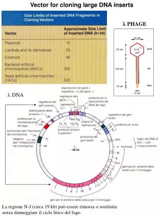
Purification and Analysis of the Mycobacterium Phage Chempanal Bryan Chempanal and Stephanie E. Simon, Department of Biological Sciences, College of Arts and Sciences, and Honors College Faculty Mentors: Lee E. Hughes, Robert C. Benjamin Department of Biological Sciences, College of Arts and Sciences BACKGROUND MATERIALS AND METHODS RESULTS From the spot test I took two of those spots and decided to branch off from there and plate each of those on separate plates and run a phage titer assay on them to determine if they were both the same phage. I named them “A” and “E”. After about two rounds of purifications I decided to continue with the “A” spot. With the “A” spot I kept running the phage titer assay until I could determine that the phage was pure. This took me about 6 rounds. After the sixth round I figured out which dilution gave me the best web pattern and name I gave this plate was “A344B-1”. I flooded this plate with 4.5 ml of phage buffer. After about two hours I filter sterilized the solution that was in the plate and ran another spot test by diluting the liquid. After performing the calculations on the spot test, I determined that the titer was 6.0 X 10^-8 pfu/ml. Next, I had to prepare a phage stock from the lysate. Using my calculation, I determined that approximately 300 pfu was needed for the web pattern and I also needed 5.0 X 10^-4 ml of phage stock to make the pattern I needed. I ended up using three times that amount to make a web pattern and decided that 15.0 X 10^-4 gave me the best web pattern. I infected 10 plates with this dilution and then put 8 ml of phage buffer in each and let them sit for about 3 hours and then aspirated them and vacuum sterilized the liquid. Next we had to run DNA preparation on the phage so that the DNA could be analyzed. The total volume of the DNA according to the nanodrop was 122.3 ug and the concentration was 165.5 ug/ul. This DNA was then used for the restriction digests which help to determine the genome size of the phage. The amount of DNA I used for the restriction digests was 4.08 ng, and this was found using the formulas that are in the National Genomics Research Initiative Phage Laboratory Manual. Using the restriction digests and the results from the gel electrophoreses, I determined that the size of the phage Chempanal was approximately 62,500 base pairs long. This was also calculated using formulas in the manual. The reason for this research is to find and isolate a mycobacterium phage and analyze its DNA to see if it is one that has been discovered yet. New discoveries will be archived and stored with its DNA for future uses. The phages used in this study were found in the soil and almost all soil contains some sort of phage. The ultimate goal of this program was to find a new phage, and that will be proved by isolating a single phage and sequencing its DNA. Methods and Materials Most of the procedures and protocols that we used throughout the research process can be found in the National Genomics Research Initiative Phage Laboratory Manual. Possibly the only variant was that we used the phage D29 as a control for most of the experiments involving infecting a host bacteria. Purification of the Phage Mycobacteria phage are mainly found in the soil, so I had to get two soil samples to purify them to extract the DNA from the phage to analyze them to see if a new phage had been discovered. The first sample I took came from the UNT campus right outside the Clark Hall, a dormitory on campus. The GPS coordinates for this this location is33.20758,-97.150973. This sample was used for a direct plating. In a direct plating sample, it is simpler and faster to get plaques than an enrichment sample but the chances of actually succeeding at a direct plating sample are pretty high. I did not succeed in my attempt because the plaques did not grow. Then I moved on to my enrichment sample. Everything I talk about from this point on will be in reference to my enrichment sample. I got my second sample from my hometown in Mesquite, Tx. There is a city recycling center I am familiar with and that is where they have a compost mill and that is where I got my sample. The GPS coordinates for this location is32.710494,-96.544966. The temperature the day I got the sample was about 85 degrees fahrenheit and the soil was slightly moist and I dug about an inch into the ground to get the sample. The protocol for the enrichment process can be found in the National Genomics Research Initiative Phage Laboratory Manual. I ended up with one plaque size and morphology, which meant that I had only one type of phage on my plate. Harvesting of a high titer lysate solution Harvesting of a high titer lysate was pretty simple once I had the plates from the final purification. I flooded the plates with phage buffer and I had to wait for about two hours before I did anything with them. The procedure for this step can be found in the National Genomics Research Initiative Phage Laboratory Manual. Basically what I had to do was to filter sterilize the solution I got from the phage buffer and phage mix and then that would have been my medium titer lysate. To find the high titer lysate required a few more calculations I had to calculate which dilution would give me the best web pattern on the plate. Once I found that, I had to plate 8 plates each with the same dilution of phage and and M.Smeg. Then once the web patterns had formed, usually in 48 hours, I had to flood the 8 plates with phage buffer and wait three hours before aspirating the liquid with a syringe and putting it in a conical tube. This is my high titer lysate. CONCLUSIONS Basically the whole point of the platings were to isolate a single type of phage from the possible many that we took up from the ground when we got our soil sample. The plaques from the phage Chempanal were medium sized, and the phage itself was an average size, measuring at about 230 nm from the end of the tail to the top of the capsid head. The capsid is about 50 nm across Because my plaques were mostly clear, it was be concluded that the phage is lytic. In lytic phage, bacterial cells are broken open and destroyed after immediate replication of the virion. As soon as the cell is destroyed, the new phages can find new hosts. As you can tell from the restriction digest picture, there are few cuts, so it was more difficult to estimate the genome size. One interesting thing about the phage Chempanal is that it is very similar to the phage Adephagia. The restriction digest images look almost identical, indicating one of two things; one, Adephagia was infected with my phage in the lab, or two, it could be a totally different phage but one very similar to Adephagia, possibly in the same family. PURPOSE AND HYPOTHESIS To understand what exactly what the drive behind this research is, one must understand what exactly a mycobacteria phage is. A mycobacteria phage is a virus that infects bacteria. Since a virus, or a phage in this case, is neither living nor dead, it must have a host to exist. In the trials that we were running in the lab, we used the host mycobacterium M. Smegmatis mainly because it is non-pathogenic and it is a fast grower, usually only requiring a 48 hour culture. Mycobacteria phages are relatively simple to manipulate and are found all over the world. Research in viral genetics is a growing field that has a lot of potential, but is relatively new. With the information that we have gained from this research, we hope to bolster an expanding library of knowledge that will continue to grow with more and more information from Universities all across the country about bacteriophages, and hopefully one day they will benefit a researcher in search of a cure for a disease. BIBLIOGRAPHY Murphy, Clare. "BBC NEWS | Health | 'Red Army' Virus to Combat MRSA." BBC News - Home. Web. 23 Nov. 2010. <http://news.bbc.co.uk/2/hi/health/6943779.stm>. "Mycobacteriophage Database | Detail for Phage Adephagia." Mycobacteriophage Database | Home. Web. 9 Dec. 2010. <http://phagesdb.org/phages/Adephagia/>. Plaques Spot Test Restriction Digest