Mycobacterium and Chlamydiae
290 likes | 314 Vues
Learn about the different entry strategies used by intracellular pathogens, such as Mycobacterium and Chlamydiae, including the "zippering" and "triggering" mechanisms. Explore the survival mechanisms of Mycobacterium and its role in the pathogenesis of tuberculosis.

Mycobacterium and Chlamydiae
E N D
Presentation Transcript
Mycobacterium and Chlamydiae Intracellular microorganisms
Bacterial Invasion Mechanism • Different strategies of entry into non-professional phagocytes have been adpoted by enteroinvasive pathogens: • the “zipper”-like mechanism – zippering of the host cell membrane around the bacterium as it enters • the “triggering” mechanism – a dramatic response at the epithelial cell surface and bacterial uptake via membrane ruffles, a process resembling macropinocytosis
“Zippering” and “Triggering” mechanisms • The zippering mechanism of entry appears to be a straightforward ligand-receptor mediated process that is characterised by cytoskeletal rearrangements that are not dramatic and disappear within a few minutes of bacterial entry. • e.g. Listeria monocytogenes entering into host cells is mediated by the internalin A – host E-cadherin interaction. • The triggering mechanism is a complex process involving many bacterial factors and an important reorganisation of the cytoskeleton at the site of bacterial entry. • e.g. Salmonella and Shigella, both secrete a set of invasion proteins via type III secretory apparatus upon contact with their cellular targets (to stimulate entry).
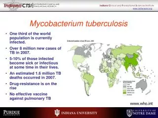
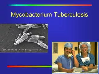
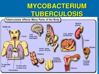
Tuberculosis • The disease is only second to HIV as the most common cause of death from a single infectious agent. • Approx 2 billion people are infected, and approx 3 million people die each year. • In developing countries, 20% of adult deaths are due to tuberculosis. • Tuberculosis is a systemic disease contracted by inhalation of M. tuberculosis-containing aerosols. • It progressively destroyed the lungs, and can spread systemically to infect any other part of the body.
Mycobacteria survive by inhibiting phagolysosome • Phagosomes with M. tuberculosis lack H+-ATPase, suggested that this bacterium prevented the late stage of endosome formation • pH of phagosome containing M. tuberculosis is 6.3-6.5 c.f. pH in most phagolysosomes. • How does M. tuberculosis control endosomal acidification? • TACO = tryptophan aspartate-containing coat protein
Role of TACO in survival of Mycobacteria Live Dead Lysosome Bacterial survival Phagolysosome
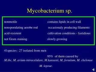
Mycobacteria • Rod-shaped, aerobic bacteria, non-sporing, slow growth rate than most bacteria. • They do not readily stain, but capable of resisting decolourisation by acid or alcohol, hence “acid-fast” bacilli. • Acid-fastness depends on the waxy envelope. • Ziehl-Neelsen technique. • Infections are acquired from inhaling infectious particles • Tubercle bacilli do not produce exotoxin or endotoxin, clinical manifestations of tuberculosis is largely from the host immune response. • Mycobacterium tuberculosis, Mycobacterium leprae, Mycobacterium avium-intracellulare.
An acid fast stain is used to diagnose the presence of mycobacteria in tissue and cytologic preparations. Note the thin red rod-like organisms (red arrows).
Identification • Culture: selective and non-selective medium. • A) Semisynthetic agar media - contain albumin, neutralises the toxic and inhibitory effects of fatty acids in the specimen. Use to observe colony morphology. • B) Inspissated egg media – plus Malachite green to inhibit other bacteria. • C) Broth media – small inocula, adding Tweens wet the media surface and permit dispersed growth of Mycobacterium. • Grow in clumps or masses because of the hydrophobic character of the cell surface.
Growth of Mycobacterium • Hydrophobic cell surface causes them to clump and inhibits permeability of nutrients into the cell.
Tubercle bacilli • Tubercle bacilli are resistant to drying and survive for long periods in dried sputum. • Mycobacterial cell walls can induce delayed hypersensitivity and some resistance to infection and can replace whole mycobacterial cells in Freund’s adjuvant. • A) Cell walls – rich in lipids, which largely bound to proteins and polysaccharides. Attribute for granuloma formation, caseous necrosis and acid-fastness.
On closer inspection, caseous necrotic tissue is seen to constitute the granulomas in this gross appearance of a Ghon complex. Most patients with primary tuberculosis are asymptomatic, and the granulomas resolve.
Tubercle bacilli • B) Proteins – when proteins bound to a wax fraction, they elicit the tuberculin reaction (sensitivity); can stimulate antibodies production. • C) Polysaccharides – role are uncertain.
Pathogenesis • Studies of Mycobacterium virulence factors and details of the bacteria-host interaction mechanism are hampered by its slow growth rate
Pathogenesis & Pathology • Proliferation of virulent organisms + interaction with the host = disease • 2 host responses: • 1) delayed-type hypersensitivity reaction to mycobacterial proteins. • 2) Cell-mediated immunity (CMI) activates macrophages to destroy mycobacteria-contained within their cytoplasm. • Tubercle bacilli spread by lymphatic channels and bloodstream. • Mycobacteria reside intracellularly in monocytes, reticuloendothelial cells, and giant cells.
Primary Infection & Reactivation • When a host has first contact with tubercle bacilli, acute exudative lesions develop. The lymph nodes undergoes massive caseation, which usually calcifies. • Reactivation is usually caused by tubercle bacilli that have survived in the primary lesion. Characterised by chronic tissue lesions, formation of tubercles, caseation and fibrosis.
The caseous necrosis is extensive, and cavitation is prominent. Such patients can be highly infectious.
Extensive cavitation of multiple granulomas of lung are typical for secondary tuberculosis from reactivation of primary infection or reinfection as an adult. Such lesions have a predilection for appearance in the upper lobes of the lung.
Tuberculin Test • Material is reactive tuberculoproteins. • Dose of tuberculin – a large amount injected intracutaneously into a hypersensitive host may give rise to severe local reactions and a flare-up of inflammation and necrosis at the main sites of infection, normally 5 TU (TU=tuberculin units). • Reactions – had primary infection with tubercle bacilli develops induration.
Diagnostic Laboratory Tests • 1) Specimens • 2) Decontamination and concentration of specimens – liquefy with N-acetyl-L-cysteine, decontaminate with NaOH. • 3) Smears - __________, __________tested for acid-fast bacilli by ________________; fluorescence microscopy with auramine-rhodamine stain. • 4) Culture : selective/non-selective; anticoagulated blood for M. avium complex; growth rate; pigmentation in the dark. • 5) DNA detection, serology, and antigen detection.
Treatment • Chemotherapy. • Tubercle bacilli prone to spontaneous mutants resistant to first-line antituberculosis drugs. • Rx: Isoniazid and rifampin. • Drug resistance to M tuberculosis is a worldwide problem.
Epidemiology • Source: human who excretes large number of tubercle bacilli, particularly from resp T; and close contact. • The development of clinical disease after infection may have a genetic component; age; undernutrition and by immunologic status; coexisting diseases (silicosis, diabetes, AIDS or HIV-infected patients).
Prevention & Control • 1) Prompt treatment, follow-up with tuberculin tests, x-rays. • 2) Drug treatment for asymptomatic tuberculin + persons. • 3) Reduced individual host resistance by starvation, gastrectomy, HIV-infected. • 4) Immunisation : BCG (bacillus Calmette-Guèrin, a live attenuated bovine organism@tubercle bacilli). • 5) The eradication of tuberculosis in cattle and the pasteurisation of milk have greatly reduced M. bovis infections.
Other Mycobacteria • Tubercle bacilli include M. tuberculosis, M. bovis. • M. avium complex commonly cause opportunistic infection ni AIDS. • M. kansaii is a photochromogen, can produce pulmonary and systemic disease identical to tuberculosis, esp in patients with impaired immune response.
Mycobacterium Leprae • Causes leprosy. • Are regularly found in skin scrapings or mucous membranes (nasal septum) in lepromatous leprosy. • Disease is divided into 2 types: lepromatous and tuberculoid. • Systemic manifestations of anemia and lymphadenopathy may also occur. Amyloidosis may develop.
Treatment and Prevention • First-line therapy is sulfones for both tuberculoid and lepromatous leprosy. • Leprosy is most likely to occur when small children are exposed for prolonged periods to heavy shedders for bacilli. Incubation period is probably 2-10 years. • Prevention is by identification and treatment of patients with leprosy, given chemoprophylactic drugs until the treatment has made them noninfectious.
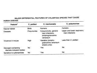
Chlamydiae • 3 species that infect human • Obligate intracellular parasites • gain energy from the host ATP • All members of Chlamydiae exhibit similar morphologic features, share common group antigen, and multiply in cytoplasm by distinctive developmental cycle.
Chlamydia trachomatis • C. trachomatis caused trachoma (an ocular infection) and genital tract infections • Exist in 2 forms: • elementary body (EB) – infectious stage • reticulate body (RB) – intracellular, non-infectious • Investigations on Chlamydia-host cell interactions is very difficult: • because it cannot be cultivated in cell-free media, • there are no well characterised mutants available, and • no genetic manipulation techniques • Adhesin.. MOMP? 40 kDa major outer membrane protein • porin enclosing a pore in the outer membrane.
Cell pathology: Elementary body EB Reticulate body RB (larger, devoid of electron-dense nucleoid) Cytoplasmic inclusion