Chlamydiae
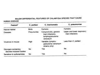
Chlamydiae. Obligate intracellular pathogens. Acute and/or persistent infections. C. trachomatis – mucosal surfaces: Ocular infections - trachoma Genital infections - pelvic inflammation, infertility Reactive arthritis C. pneumoniae Pneumonia Atherosclerosis. Chlamydia Life Cycle.

Chlamydiae
E N D
Presentation Transcript
Chlamydiae • Obligate intracellular pathogens. • Acute and/or persistent infections. • C. trachomatis – mucosal surfaces: Ocular infections - trachoma Genital infections - pelvic inflammation, infertility Reactive arthritis • C. pneumoniae Pneumonia Atherosclerosis
Chlamydia Life Cycle • Elementary body (EB) -metabolically inactive -highly infective stage • Reticulate body (RB) -metabolically active -intracellular growth stage • Persistent body (PB) -life cycle pause between EB and RB stages -stable association with host cell
Apoptosis vs Necrosis • Apoptosis – programmed cell death. -eliminate and phagocytose cells in an orderly fashion. -phosphatidylserine (PS) receptor on phagocytes increases anti-inflammatory cytokines TGF-beta and IL-10. -needed for embryogenesis, immune system maintenance. • Necrosis – non-programmed cell death. -cellular debris is a ‘danger signal’ in the cell, so inflammatory response follows DSR interaction with phagocytic cells.
How might apoptosis help pathogens? • Facilitating pathogen propagation -pathogens within apoptotic cells can be taken up by other phagocytic cells without the pathogen having to navigate the extracellular environment. • Avoiding inflammatory responses -apoptosis can release anti-inflammatory cytokines that down regulate the immune response.
Pathogen manipulation of apoptosis • Viruses – often inhibit apoptosis • Oncogenic viruses destroys p53 surveillance system • Inhibit extrinsic and intrinsic apoptosis pathways • Protozoa – often inhibit apoptosis • Toxoplasma, Trypanosomes, Cryptosporidium • Heat shock proteins, NF-KB • Bacteria – often induce apoptosis - Helicobacter, Shigella, Salmonella - Toxins, protein synthesis inhibitors, TTSS
Mechanisms of Apoptosis • Extrinsic pathway – Receptor mediated • FasL-death receptor interactions • Initiator caspases – caspases 8, 9 • Effector caspases – caspases 3, 6, 7 • Intrinsic pathway – Intracellular origin • Caspase activation or intracellular stress signals • Mitochondrial release of cytochrome c • Apoptosome formation
Extrinsic pathway • Type I cells – activate initiator caspase 8, then effector caspase 3, then apoptosis commenses. • Type II cells – require mitochondrial amplification. BAX, BAK stop being inhibited by BCL-2, BCL-X and cause mitochondrial release of cytochrome c, then apoptosome forms, activates caspase 3, and commenses apoptosis (DNA fragmentation, nuclear condensation, membrane blebbing, etc.)
Chlamydial apoptosis manipulation • When to inhibit apoptosis? • When to induce apoptosis?
Chlamydial apoptosis manipulation • When to inhibit apoptosis? - for chronic or persistent infections - when intracellular growth stages dominate • When to induce apoptosis? • for acute infections • when infectious Elementary Body stages dominate
How to manipulate apoptosis? • ‘Chlamydia protein associating with death domains’ = CADD, is an oxidoreductase, so accumulation of reactive oxygen species could lead to necrosis, while interactions with Fas could inhibit apoptosis. • Chlamydia interferes with mitochondrial apoptosis signals, perhaps by secreting Bcl-2 anti-apoptotic proteins or inactivating pro-apoptotic proteins. Type III Secretion Systems available.
How to induce apoptosis? • Caspase-independent apoptosis occurs. • Necrosis occurs in some cases… by design or accident? • Cell type specific interactions. • Over-expression of BAX, BAK cause cell death. • BAX deficient cells and mice had fewer Chlamydial organisms, so perhaps BAX-induced apoptosis is important for propagation of the infection. • Increased mitochondrial metabolism and oxidative stress observed in infected cells.
How to inhibit apoptosis? • Inhibit cytochrome c release from mitochondria. • MEK/ERK – MAPK signalling pathways. • NF-KB – as with MEK/ERK, upregulate transcription of anti-apoptotic genes. • IAP – upregulate Inhibitors of Apoptosis Proteins.
Future Work No methods exist for genetic transfer (yet), so much is unknown about virulence and pathogenesis! Is Chlamydiae-induced apoptosis associated with acute disease while Chlamydiae-inhibited apoptosis is associated with chronic disease?