Download
1 / 20
240 likes | 618 Vues
Prenatal Diagnosis of Congenital Malformations. Max Brinsmead PhD FRANZCOG January 2010. 3 categories of congenital disorder. Some 1- 2% of babies will have a major disability that dates from the prenatal period Either Chromosomal disorder e.g. Down syndrome

E N D
Prenatal Diagnosis of Congenital Malformations Max Brinsmead PhD FRANZCOG January 2010
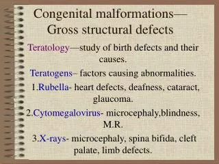
3 categories of congenital disorder • Some 1- 2% of babies will have a major disability that dates from the prenatal period • Either • Chromosomal disorder e.g. Down syndrome • Structural abnormality e.g. “Hole in the Heart” • Other e.g. Cerebral palsy • While a good deal of antenatal care in the 21st century is directed to the prenatal detection of these problems.... • It is worth remembering that, for most, termination of the pregnancy is the only option
What can be detected in utero? • All chromosomal abnormalities • About half of the major structural abnormalities • May be difficult in the 1st half of pregnancy • And varies with the resources available • Very few of the patients with cerebral palsy
Types of Tests • Diagnostic tests • Have a high degree of accuracy • e.g. Amniocentesis for chromosomes • May be invasive, carry risk and expensive • Screening tests • Cheap and safe tests suitable for whole population • Selects a subgroup for diagnostic testing • So sensitivity and positive predictive value is important • Ultrasound can be used for both screening and diagnosis • Limited only by the resources available
Two Practical Characteristics of a Screening Test • Sensitivity • The number of patients selected by the test as positive as a proportion of the total number of patients with the condition sought • e.g. A DRA test that identifies 3 out of 4 mothers with a Down syndrome baby has a sensitivity of 75% and will therefore miss 25% of the babies with this problem • Positive Predictive Value (PPV) • The likelihood that a patient identified by the test has the condition sought • e.g. A DRA with PPV of 10% means that 90% of women selected to undergo amniocentesis will NOT have this condition
Markers for Down Syndrome • Ultrasound • Nuchal translucency (NT) ↑with DS • Nasal bone (absent with DS) • Maternal Serum • PAPP-A ↓with DS • Beta HCG ↑with DS • AFP ↓with DS and ↑with NTDs • Oestriol ↓with DS • Inhibin-A (useful in 2nd trimester testing)
Down Syndrome screening tests • Use the combined test (i.e NT measure, beta-HCG & PAPP-A )for patients who present in the first trimester • Sensitivity when optimally timed is 85-88% • Can help many patients avoid amnio & CVS • Use the Triple Test (i.e. beta-HCG, E3 and AFP) for patients who present in the 2nd trimester • Sensitivity when optimally timed is 65 – 75% • Also screens for NTDs • Integrated testing offers greater sensitivity and higher PPV but results are late
Information required for the interpretation of tests • Gestational age with accuracy • And this is why ultrasound c NT is so useful • Maternal age • Advancing age will increase the chance of a +ve result • Maternal ethnicity and weight • Multiple pregnancy? • Assisted conception? • Maternal diabetes? • Maternal smoking?
Timing is all important • Firstly it is desirable to make a diagnosis before 14w so that TOP is simple and safe • 1st blood test between 9 – 13w&6d • Optimally 10 -12 w • NT measure at 11 – 13w&6d • Optimally at 11.5 – 13.5w • Second trimester blood test 14 – 20w • Optimally 14 – 18w • CVS can be performed any time after 9.5w • Amniocentesis after 15w
Timing is all important • Full chromosomal analysis from CVS or amnio takes 10 – 14d • Culture failure occurs in <0.5% • Colony mosaicism can be a problem • Some abnormalities can pose dilemmas • e.g. 47 XYY, 47XXY, 45XO • Fast FISH or DNA tests can be done overnight • But are much more expensive • And have a small risk of error
Some myths surrounding PND • Most Down Syndrome babies are born to women over the age of 35 • Because my other pregnancies and babies were normal I do not need testing • There is no Family History of Downs so I do not need testing • Husband’s age is important • The chance of aneuploidy after a positive screen test is equivalent to the risk calculated
Amniocentesis and CVS • Practitioners of both state that the increased loss associated with both is about 1:100 • Large studies say 0.5% for amnio and 1-2% for CVS • Most pregnancy losses occur within 7 – 10d • A few patients leak liquor after amnio • CVS is done PA for an anterior placenta and PV for a posterior placenta • Both procedures require US guidance • And are best done by practitioners of >50/yr
Issues a patient needs to understand before undergoing a test • What the test is for • And what it will not detect • What is their risk of the condition • What is the likelihood that the test will be positive • What are the sensitivity and PPV of the test • What will they do if the test is positive • What are the risks associated with further testing. What will that test reveal • What will they do if their baby is affected
So Patient Counselling is Important • Pre test counselling will help to reduce post positive test anxiety • Resources are required for the optimal management of screen-positive patients • And the continuing care of patients who are screen-positive but amnio/CVS normal • Because they do have pregnancies at increased risk • Consultation with a MFM specialist may be desirable
Increased Nuchal Translucency • When aneuploidy has been excluded... • Risk of miscarriage <20w is 1.3% for NT 95 – 99th centile • But is 20% when NT >6.5 mm • Look for cardiac abnormalities • 1.7% if NT is 2.5 – 3.4 mm • 7.5% if NT >3.5 mm • Is it Cystic Hygroma? i.e. generalised oedema • 5-fold increased risk of aneuploidy • 12-fold increased risk of cardiac problem • 6-fold increased risk of perinatal death • So monitor – consult MFM if it does not resolve
Low PAPP-A • When beta-HCG is high exclude Downs • (Trisomy 13 & 18 associated with low beta HCG) • When aneuploidy is excluded.... • Monitor for early onset pre eclampsia & IUGR • ? Consult with MFM especially if beta HCG is high • Unexplained elevation of PAPP-A • “Not associated with any known pregnancy problem”
Raised AFP • Causes include: • Wrong dates • Pregnancy bleeding • Multiple pregnancy • Open neural tube defects* • Anterior abdominal wall defects • Some rare tumours • So refer for tertiary level scanning • and • “Watch carefully” for the rest of the pregnancy
Low oestriol in the 2nd Trimester • Think about: • Placental sulfatase deficiency • X-linked recessive • Ichthyosis (lizard-like skin) • Mild corneal opacification • Cryptorchidism • Smith Lemli-Opitz Syndrome (rare 1:50,000) • Multiple malformtions • Mental retardation • Syndactly • So consider amniocentesis or CVS