Download

1 / 83
1.08k likes | 2.66k Vues
Radiology of The Ear. MRI. Plain X Ray. CT Scan. Demonstrates VIII nerve Brain Great vessels. Of limited value Demonstrates Mastoid air cells. Accurately demonstrates External ear Middle ear Surrounding structures. lateral Oblique (Mastoid)view.

E N D
Radiology of The Ear MRI Plain X Ray CT Scan Demonstrates VIII nerve Brain Great vessels Oflimited value Demonstrates Mastoid air cells Accurately demonstrates • External ear • Middle ear • Surrounding structures
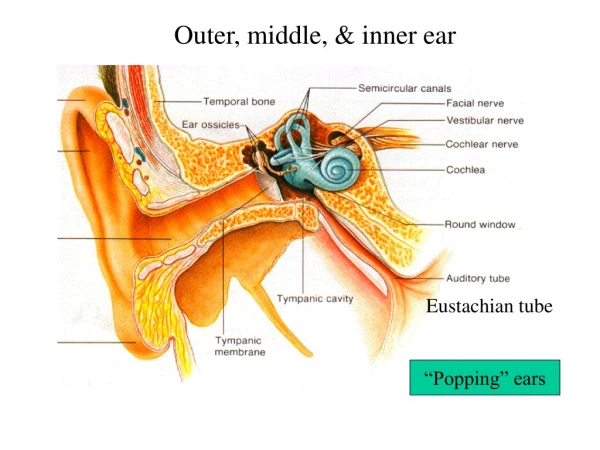
lateralOblique (Mastoid)view • Locate the Temporo-mandibular joint the external auditory canal (EAC) which is a complete circle • The mastoid air cells are behind and above the EAC
lateralOblique (Mastoid) TMJ External auditory canal Pneumatised mastoid: air spaces separated by bony partitions
The mastoid cells (white arrow) are obscured, and not air-containing, due to chronic otitis media. External auditory canal TMJ
Schüller view: Well-developed normally pneumatized mastoid air cells can be observed in the picture on the left side (double arrow). • In the picture on the right side, the mastoid cells (arrow) are obscured, and not air-containing, due to chronic otitis media. TMJ Sinodural angle EAC
External auditory canal TMJ • There is a clean cavity behind and above the external auditiry canal not surrounded by sclerosis • Diagnosis: surgical cavity of mastoidectomy
Axial CT scan, the destructed apex of the petrous bone can be observed (white arrow), which is caused by ? cholesteatoma. Petrous bone
Axia CT scans: • The mastoid cells on the right side (green arrow) are totally obscured, which proves mastoiditis. • On the left side (blue arrow), an intact status can be seen.
Axial CT scans: Transverse temporal bone fracture (arrows).
imaging of the nose MRI Plain X Ray CT Scan Accurately demonstrates • Nose • Paranasal sinuses • Surrounding structures Mainly for Surrounding soft tissue structures limited value Screening of sinuses Medico-legal IN NASAL BONE FRACTURE
Occipito-mental projection • Patient facing the film • Radiologic base line tilted 450 • Beam horizontal , directed to external occipital protuberance
Frontal sinus orbit septum Maxillary sinus Maxillary sinus Sphenoid sinus
NB • Radiologic Examination of sinuses should be: • In erect position • Sphenoid is seen in occipitomental view with open mouth
Frontal Sinus Ethmoid Sinus Maxillary Sinus Soft Palate Nasopharynx Sphenoid Sinus Sella Turcica Clinoid Process
Occipito-mental view of the sinuses showing partial opacification of the right maxillary sinus, with an air-fluid level AcuteSinusitis
An air-fluid level AcuteSinusitis
Loss of continuity of nasal bone with displacement of distal fragment NASAL FRACTURE
Axial view Coronal view CT SCAN
Coronal CT scanNormal findings • The sinuses normally contain air which is seen in black color • The frontal sinus : • Above the orbit • Seen in the anterior cuts • May be absent ORBIT ORBIT
Ethmoid sinuses • 15 to 20 air cells in each side • Medial to Lamina paparycea Maxillary sinus - Below the orbit ORBIT Ethmoid Maxillary Maxillary
Sphenoid Sinus • Divided by a septum into right and left sinuses • The floor of the sinus is the roof of the nasopharynx Sphenoid
Orbit Bulla Ethmoidalis Middle Turbinate Maxillary Sinus Middle Meatus Uncinate process Inferior Turbinate Inferior Meatus
CORONAL CT SHOWING THICKENING OF THE FRONTAL SINUS MUCOSA
Osteoma. A left frontal osteoma ( arrow) is visible anteriorly in this coronal CT scan. Note its increased density, characteristic of the lesion.
Coronal CT scan showing normal ostiomeatal complex. Patent ostia are visible on both sides, and sinuses are well ventilated.
Coronal CT scan • Total ethmoid opacity ( ethmoidal polypi) • Fluid level in the left maxillary sinus • Diagnosis : bilateral ethmoid sinusitis Left maxillary sinusitis
Coronal CT scan • Blocked osteomeatal complex • Opacity of right ethmoidal air cells • Fluid level in the left maxillary sinus • Thickened mucosa of right maxillary sinus • Diagnosis: bilateral Maxillary sinusitis, right ethmoid sinusitis
Coronal CT scan Blocked ostiomeatal complex Maxillary sinus Maxillary sinus
A coronal CT scan • Moderate bilateral maxillary sinus mucosal thickening with blockage of both ostiomeatal complexes • Chronic sinusitis
A coronal CT scan. • Complete opacification of the right maxillary sinus • Mucosal thickening of the left maxillary sinus • Chronic sinusitis
Coronal CT scan • Concha bullosa i.e pneumatized middle turbinate • A deviated nasal septum.
Concha bullosa i.e pneumatized middle turbinate ( red arrow). orbit orbit Maxillary sinus
Coronal CT scan Bilateral total opacity of ethmoid sinuses Bilateral Ethmoidal polypi
Coronal CT scan showing right maxillary sinus opacification. Also, note the septal deviation to the right and the hypertrophy of the left inferior turbinate (yellow arrow)
Coronal CT scan of the sinuses showing bilateral maxillary sinusitis. The opacification is more prominent on the left side (arrow).
Oroantral fistula Enumerate 3 causes starting with the most common cause
Complete right maxillary sinus opacity • Opacity and Widening of the right osteomeatal complex • Soft tissue opacity in the nasopharynx
Soft tissue mass in the nasal cavity and left maxillary and ethmoidal sinuses The left middle meatus and medial wall of the left maxillary sinus are absent. There is mucosal thickening of the right maxillary sinus Differential Diagnosis Inverted papilloma Antrochoanal polyp Squamous cell carcinoma Inverted Papilloma
Coronal CT scan • Bilateral sphenoidal sinus opacity • Diagnosis: Bilateral Sphenoid sinusitis
There is soft-tissue thickening over the expanded Right Frontal Sinus ?? left Frontal sinuses are partially opacified by mucoperiosteal thickening Axial CT scan expansion of the Right Frontal sinus.
Hyperdense sinus secretions. This axial CT scan shows hyperdense secretions in the left maxillary antrum. fungal sinusitis.
Sinonasal polyposis. Note the polypoid changes with opacification and expansion of the right Nasal cavity, right maxillary sinusitis coexists.
MRI • Coronal MRI scan showing opacification of the left maxillary and ethmoid sinuses
Axial MRI scan showing opacification of the left maxillary sinus
Barium swallow Plain X Ray CT Scan Accurately demonstrates • Pharynx • Surrounding srtucture • with LN The lumen ++ limited value demonstrates Lumen of pharynx