Download
1 / 25
290 likes | 744 Vues
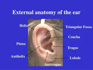
Anatomy of The Ear. Anatomy of The Ear. EXTERNAL EAR MIDDLE EAR INNER EAR. Helix. Crus of Helix. The external Ear 1- The Auricle 2-The External Auditory Canal 3-The Tympanic Membrane. Antihelix. Tragus. Concha. Lobule. SITE Atached to the side of the head SHAPE

E N D
Anatomy of The Ear • EXTERNAL EAR • MIDDLE EAR • INNER EAR
Helix Crus of Helix The external Ear 1-The Auricle 2-The External Auditory Canal 3-The Tympanic Membrane Antihelix Tragus Concha Lobule SITE Atached to the side of the head SHAPE Oval & Flat, with series of ridges and depressions on lateral surface STRUCTURE Plate of fibroelastic cartilage covered by skin which is closely adherent to the underlying perichondrium Areas devoid of cartilage: 1- lobule Incisura terminalis
Extends from concha to the tympanic membrane Length: 24 mm Outer 1/3: cartilaginous Inner 2/3 bony The external Ear • The Auricle • The External Auditory Canal • The Tympanic Membrane
Outer 1/3: directed upward, backward and medially Skin is - thick - Contain hairs, sebaceous and ceruminous glands Inner 2/3: directed downwards, forwards and medially Skin is thin , closely adherent to underlying perichondrium No hairs or glands
The external Ear The Auricle The External Auditory Canal The Tympanic Membrane • Shape; • oval, about 1 cm in diameter • Concave medially • Two unequal pars: • 1- Pars flaccida (small upper part ) 2- Pars tensa (major lower part) • Site : - At the medial part of the external canal - Separates the external ear from the middle ear
Structure of the tympanic membrane THREE LAYERS 1-Outer skin layer; contineous with the skin of the external canal 2- Middle fibrous layer: well formed in Pars tensa while poorly formed in Pars flaccida 3 Inner mucosal layer : contineous with the mucosa of the middle ear
The Middle Ear Cleft: 1-Middle Ear 2-Eustachian Tube 3-Mastoid Air Cells
The Middle Ear Cleft: 1-Middle Ear SITE -between the external & Middle Ear SHAPE Like biconcave lens Height: 15mm Antro-posteriorly: 15mm From side to side : 2mm
The Middle Ear Cleft: 1-Middle Ear Structure: All the walls are bony except the lateral wall (Tympanic membrane) Lining Epithelium; • Antro-inferiorly, ciliated columner epithelium • Postero-superiorly: flattened squamous epithelium
The Middle Ear Cleft: 1-Middle Ear Contents: • Air • Three ossicles • Two muscles • One nerve
The Middle EarCleft: 1-Middle Ear Compartments 1-Mesotympanum Opposite the tympanic membrane 2-Epitympanum Above the tympanic membrane 3-Hypotympanum Belowthe tympanic membrane
Walls of the Middle Ear مكعب له ستة أوجه Lateral wall Medial wall Superior wall Inferior wall Anterior wall Posterior wall -Separates the external ear from the middle ear -Formed mainly of tympanic membrane with small bone above and below the drum
Walls of the ME Lateral wall Medial wall Separates the ME from The Inner Ear Promontory; formed by basal turn of cochlea Oval window: above &behind the promontory, closed by footplate of stapes Round window: below & behind the promontory, closed by secondary tympanic membrane Horizontal part of VII nerve: above the promontory & oval window
Walls of the ME • Lateral wall • Medial wall • Superior wall Called Tegmen Tympani Thin bony plate Separate the ME from the Temporal lobe
Walls of the ME • Lateral wall • Medial wall • Superior wall • Inferior wall Thin bony plate Separate the ME from the Jagular Bulb
Walls of the ME Lateral wall Medial wall Superior wall Inferior wall Anterior wall Separates the ME from the ICA Tensor Tympani muscle enter the ME through this wall Has an opening for ET
Walls of the ME • Lateral wall • Medial wall • Superior wall • Inferior wall • Anterior wall • Posterior wall - Separates ME from Mastoid process - An opening: Aditus ad antrum : connects the epitympanum with the mastoid antrum - The stapedius muscle enters the ME through this wall - The vertical part of VII nerve runs through this wall
The Eustachian Tube - connects the ME with the Nasopharynx • Directed downward forwards and medially • Length: 36 mm. • It is closed at rest and opens during swallowing yawing by contraction of tensor palati muscle • Its lateral 1/3 is bony & medial 2/3 is cartilagenous • In children it is shorter, more horizontal & wider
The Mastoid Air Cells Within the mastoid process Pyramidal Filled by air cavities ie pneumtized The largest is the antrum Pneumatization varies: 1- Pneumatic mastoid: highly cellular (the commonest) 2-Diploic mastoid: less cellular 3- Sclerotic: contains only few cells The cells are lined by flattened squamous epithelium
The Inner Ear • Within the temporal bone • Between the Middle Ear & Internal Auditory Canal
The Bony Labyrinth Hollow bony capsule Oval window & round window opens into it Consists of 1- bony Cochlea 2- Bony semicircular canals 3-vestbule
Membranous Labyrinth Within the bony cochlea Consisits of 1- cochlear duct in the bony cochlea 2- semicircular ducts within the bony semi-circular canals 3- saccule & utricle in the vestibule 4-Endolymphatic duct & sac
The membranous labyrinth is filled with endolymph • Surrounded by perilymph
The sensory end organs: • In the Cochlea : Organ of Corti • In The Semi-circular duct: The Crista • In the Utricle & Saccule The Macula