Download

1 / 46
590 likes | 2.15k Vues
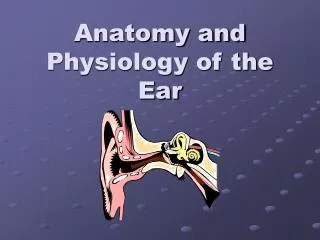
Anatomy and Physiology of the Ear. The Temporal Bone Outer Ear Middle Ear Inner Ear Cochlear Physiology. Which Way?. Anterior/Ventral = toward the front Posterior/Dorsal = toward the back Lateral = toward the side Medial = toward midline

E N D
Anatomy and Physiology of the Ear • The Temporal Bone • Outer Ear • Middle Ear • Inner Ear • Cochlear Physiology
Which Way? • Anterior/Ventral = toward the front • Posterior/Dorsal = toward the back • Lateral = toward the side • Medial = toward midline • Superior = toward upper surface (rostral) • Inferior = toward lower surface (caudal)
Gotta Catch a Plane Sagittal- dividing right from left Coronal(Frontal) -dividing front from back Horizontal-dividing up from down
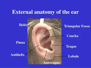
The Outer Ear Consists of: • The Pinna - cartilaginous, highly variable in appearance, some landmarks. • External Auditory Canal (or external auditory meatus) - 2.5 cm tube.
Pinna Landmarks • Helix • Antihelix • Concha • Tragus • Intertragal Notch • Antitragus
External Auditory Canal • lateral portion-cartilage • medial portion-osseous • lined with epidermal (skin) tissue • hairs in lateral part • cerumen (ear wax) secreted in lateral part.
Outer Ear Functions 1 • Amplification / Filtering -- increases sounds between 1500 and 7000 Hz by 10 to 15 dB -- because of the resonance of Concha -- 5000 Hz E.A.Canal -- 2500 Hz
Outer Ear Functions 2 • Protection -- medial displacement of ear drum -- curvature of canal -- hairs -- cerumen -- skin migration
Outer Ear Functions 3 • Localization -- The ability to identify the location of a sound source -- (Will be covered more later)
The Middle Ear:A cleft within the temporal bone • Lining is mucous membrane • Tympanic Membrane separates it from EAC • Eustachian tube connects it to nasopharynx • Also Connected to Mastoid Air Cells
Middle Ear Structures 1- Malleus 2- Incus --Ossicles 3- Stapes 4- Tympanic Membrane (Eardrum) 5- Round Window 6- Eustachian Tube
Middle Ear Muscles1. The Stapedius Attaches to Stapes Contracts in Response to Loud sounds, chewing, speaking Innervated by the Facial (VIIth cranial) nerve
Middle Ear Functions • Impedance Matching -- amplification of sounds to overcome difference in impedance between the air of EAC and the fluid of the inner ear. • Filtering -- resonant frequency is approximately 1000 Hz, functions as bandpass filter. • Acoustic Reflex -- Contraction of Stapedius muscle in response to loud sounds
Middle Ear Function • Impedance Matching is accomplished through pressure increase produced by the middle ear. • From 2 main effects: Reduction in AREA Increase in FORCE
Reduction in AREA • sound striking the (relatively large) tympanic membrane • is delivered to the (much smaller) stapes footplate • Areal Ratio = 18.6 to 1
Increase in FORCE • The malleus and incus act like a lever • Whenever there is a pivot: • Force x Length in = Force x Length out • Force is greater on short side (Think of wheeled luggage) • Malleus manubrium = 1.3 times as long as Incus long process
Leverage • Small force (baby’s weight) supports man • because of the difference in length on either side of the pivot point
Increase in Pressure • Remember that Press. = Force/Area • force is increased 1.3 times • area is decreased 18.6 times • Pressure is increased 24.2 times (27.7 dB)
Other Key Middle Ear Function • Oval Window Isolation-- Sound striking the tympanic membrane is delivered through the ossicular chain to the oval window • Without the middle ear, both the oval and round windows would receive sound energy and energy would cancel out.
Middle Ear Filtering: • Band Pass filter • Resonant Frequency near 1kHz • Effect can be seen in Minimum Audibility Curve (Figure 10.2)
Minimum Audibility Curve (Figure 10.2) • Plot of threshold of detection (in dB SPL) for tones as a function of frequency. • Shows: best hearing around 1 kHz poorer hearing below 500 Hz and above 4000 Hz
Tympanometry • Acoustic measures of middle ear health • Made using an immittance (or impedance) bridge: • PRESSURE PUMP/MANOMETER • MINIATURE SPEAKER • MICROPHONE • ALL CONNECTED THROUGH A SMALL PROBE INSERTED IN EAR CANAL
Compliance: opposite of stiffness. • middle ear system is not massive, largely a stiffness-controlled system. • Changes in stiffness/compliance have large effects on functioning of system. • at point where air pressure in canal and middle ear are equal the most sound will be conducted through.
Tympanogram: • A plot of middle ear compliance as a function of ear canal pressure • Pressure is swept from +200 to -200 or -400 dPa • Should see peak at point where pressures are equal
Tympanogram types: • A: peak between +100 and -200 dPa: normal • C: peak beyond -200 dPa: neg pressure • B: no peak flat tymp: effusion • As: peak but shallow: stiff: otosclerosis • Ad: peak off scale: floppy: dysarticulation
The Acoustic Reflex • Stapedius contraction measured as change in compliance • Reflex arc: • peripheral ear, • VIIIth n. • Cochlear nucleus • superior olivary complex • VIIth n. to the middle ear • Reflex is bilateral.
Clinical Tests using Acoustic Reflexes: • A.R. Threshold: how intense sound must be to elicit the reflex? • A.R. Decay: Is the degree of a contraction maintained throughout a 10 second stimulus?
INNER EAR Two Halves: • Vestibular--transduces motion and pull of gravity • Cochlear--transduces sound energy (Both use Hair Cells)
Subdivision into spaces containing endolymph (blue), and spaces containing perilymph (red)
Cochlea is Divided into 3 “Scala” • Scala Vestibuli • Reissner’s Membrane • Scala Media • Basilar Membrane • Scala Tympani • Helicotrema - the opening between 2 outer Scala
Fluids filling the Inner Ear • Perilymph- in S. Vestibuli and S. Tympani • High Sodium / Low Potassium concentrations • Low Voltage (0 to +5 mV) • Endolymph- in S. Media • High Potassium / Low Sodium concentrations • High Positive Voltage (85 mV)
Cross-Section of the Cochlea Third Turn Second Turn First Turn
I = Inner Hair Cells P = Pillar Cells O = Outer Hair Cells D = Deiter’s Cells
The Stereocilia on IHCs and OHCs • OHCs (at top) • V or W shaped ranks • IHC (at bottom) • straight line ranks
Cochlear Functions • Transduction- Converting acoustical-mechanical energy into electro-chemical energy. • Frequency Analysis-Breaking sound up into its component frequencies
Transduction- • Inner Hair Cells are the true sensory transducers, converting motion of stereocilia into neurotransmitter release. Mechanical Electro-chemical • Outer Hair Cells have both forward and reverse transduction-- Mechanical Electro-chemical Mechanical Electro-chemical
Frequency Analysis-the Traveling Wave Bekesy studied cochleae from cadavers, developed the Traveling Wave theory 1. Response always begins at the base 2. Amplitude grows as it travels apically 3. Reaches a peak at a point determined by frequency of the sound 4. Vibration then dies out rapidly