Digestive System
Digestive System. Chapter 14. Introduction to Digestive System. Converts food into raw materials that build and fuel body’s cells Ingestion Digestion Absorption Defecation. Anatomy of Digestive System. 2 groups Alimentary canal (GI Tract) Ingestion, digestion, absorption, defecation

Digestive System
E N D
Presentation Transcript
Digestive System Chapter 14
Introduction to Digestive System • Converts food into raw materials that build and fuel body’s cells • Ingestion • Digestion • Absorption • Defecation
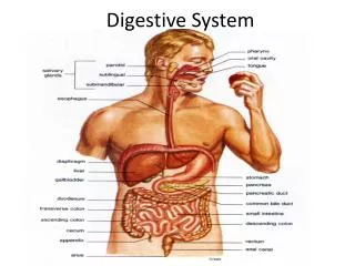
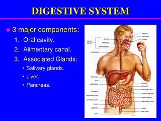
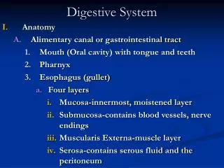
Anatomy of Digestive System • 2 groups • Alimentary canal (GI Tract) • Ingestion, digestion, absorption, defecation • Continuous, coiled muscular tube through body • Open at both ends • Food outside body • Accessory digestive organs • Assist digestive breakdown Figure 14.1
Organs Alimentary CanalMouth • Entry of food, oral cavity • Lips • Cheeks • Hard Palate, Soft Palate • Vestibule • Tongue • 2 bony attachments – Hyoid and styloid process of skull • Lingual frenulum – mucous membrane secures tongue • Mastication – chewing • Mixing of food with saliva, tongue moves around, food breakdown begins before leaving mouth Figure 14.2
Organs of Alimentary CanalPharynx • Only Oropharynx and laryngopharynx • Walls contain two muscles • Inner longitudinal • Outer circular • Alternating action = peristalsis • Move food downward Figure 13.2
Organs of Alimentary CanalEsophagus • Runs from pharynx through diaphragm to stomach • Conduct food by peristalsis to stomach Figure 14.1
Walls of Alimentary CanalEsophagus to Large Intestine • 4 basic tissue layers • Mucosa – lines lumen, epithelium and connective tissue • Submucosa – connective tissue layer, contains blood vessels, nerve endings, lymph nodules, lymphatic vessels • Muscularis externa – circular and longitudinal smooth muscle • Serosa – serous fluid-producing cells form visceral peritoneum, continuous with parietal peritoneum by way of mesentary Figure 14.3
Organs of Alimentary CanalStomach Figure 14.4a • C shaped left side • Greater, Lesser Curvature • Regions • Cardiac – surrounds cardioesophageal sphincter • Fundus • Body • Pyloric Antrum • Pylorus – continuous with small intestine through pyloric sphincter Figure 14.4b
Omentum and Mesenteries • Lesser omentum – double layer peritoneum, liver to lesser curvature • Greater omentum – double layer peritoneum, drapes, covers organs, attaches to cavity wall • Cushions, insulates, protects, lymph nodules, macrophages, immune cells Figure 14.5
Stomach • Circular, longitudinal and oblique layer of muscle – muscularis externa • “storage tank” • Initiation of protein breakdown • Cells that line bicarbonate rich mucus – protects from HCl and digestive enzymes Figure 14.4a
Cells of Stomach • Lining dotted with gastric pits -> gastric glands • Chief cells – protein digesting enzymes - pepsinogens • Parietal cells – HCl, activates enzymes • Enteroendocrine cells – local hormones, digestive activity of stomach – gastrin • Most digestion occurs in pyloric region • After process, food looks like heavy cream = chyme
Peptic Ulcers – How stressed are you? • Burning pain 1-3 hrs after eating, loss of appetite, bloating, nausea, vomiting • 1 in 8 • Most occur in pyloric region of stomach or duodenum • Erosion due to exposure to HCl • Stress? Can aggravate it • NO! Helicobacter pylori • 90-100% duodenal • 80-90% gastric
Organs of Alimentary CanalSmall Intestine • MAJOR digestive organ • 3 subdivision • Duodenum • Jejunum • Ileum – meets large intestine at ileocecal valve • Only able to process small amount of food at time, pyloric sphincter controls food movement
Accessory Organs Associated with Duodenum • Pancreas – enzymes that complete food digestion, ducted to duodenum via pancreatic ducts • Gallbladder and liver – bile(formed by liver) enters via bile duct • Bile duct and pancreatic duct form hepatopancreatic ampulla and duodenal papilla Figure 14.6
Walls of Small Intestine • Nearly all food absorption • 3 structures in wall increase absorptive surface • Microvilli – plasma membrane – brush border enzymes – digestion of protein and carbs • Villi – mucosa, w/in each are capillaries and lacteal duct, food absorbed • Circular folds – deep fold of mucosa and submucosa, don’t disappear as fill with food • All decrease as move along • Peyer’s Patches increase – collections of lymphatic tissue • Bacteria in food! Figure 14.7
Organs of Alimentary CanalLarge Intestine • Ileocecal valve to anus • Absorb water and eliminate indigestible food as feces • No villi • Goblet cells in mucosa produce HCO-3 rich mucus, lubricant • Divisions • Cecum, Appendix, Colon, Rectum, Anal Canal • Colon – ascending, transverse, descending, sigmoid • Anus – internal and external sphincters Figure 14.8
Accessory Digestive OrgansTeeth • Masticate – chew • Teeth – tear and grind breaking down into smaller fragments • Deciduous Teeth • 6 months – 2 yrs (20 teeth) • Permanent Teeth • 6-12 yrs roots of baby teeth reabsorbed and fall out • Wisdom teeth emerge by 17-25 if going to Figure 14.9
Accessory Digestive OrgansSalivary Glands • Three pairs of glands • Parotid gland – Mumps • Submandibular and Sublingual • Empty secretions into floor of mouth via ducts • Produce Saliva – mucus and serous fluid • Mucus moistens, bind = bolus • Serous contains – salivary amylase in bicarbonate solution, begins digestion of starch in mouth • Lysozyme and IgA • Dissolves food chemicals to be tasted
Accessory Digestive OrgansPancreas • Only organ produces enzymes that break down all categories of digestible food • Secreted into duodenum in alkaline fluid • Why alkaline? • Endocrine function
Accessory Digestive OrgansLiver • One of body’s most important organs and largest gland • Four lobes, suspended from diaphragm and abd. wall by falciform ligament • Digestive function – produce bile
Bile • Yellow-green color • Watery solution contain bile salts and pigments, cholesterol, phospholipids, electrolytes • Bilirubin – breakdown product of Hb • Only bile salts and phospholipids aid digestion – emulsify fats, more SA for fat digesting enzymes to work on
Accessory Digestive OrgansGall Bladder • Thin walled green sac • When not eating bile stored here • Concentrated by water removal • Fatty meal enters duodenum, bile released due to hormonal stimulus
Gallstones • Bile stored for too long or too much water removed, cholesterol crystallizes • Blockage of common hepatic or bile ducts prevent bile into small intestine, backs up into liver • Bile pigments enter blood and circulate, yellowing jaundice http://gogogojiteam.com/gallstone.html
Functions of the Digestive SystemIngestion • Food must enter the system to be acted upon • Active and voluntary Figure 14.11
Functions of the Digestive SystemPropulsion • Foods must be processed my multiple organs – move from one to next • Swallowing is example • Movement depends mostly on peristalsis vs. segmentation • Segmentation – small intestine, alternating segments contract, mechanical digestion
Functions of the Digestive SystemFood Breakdown: mechanical digestion • Mixing of food in mouth by tongue • Churning of food in stomach • Segmentation in small intestine • Prepare food for enzymatic digestion by breaking into smaller pieces
Functions of the Digestive SystemFood Breakdown: chemical digestion • Food molecules broken down into building blocks by enzymes • Reactions called hydrolysis rxn, water added as bond breaks • Carbs (saccharides) we digest – sucrose, lactose, maltose, and starch. Eat cellulose but can’t digest, fiber • Proteins (a.a.) – polypeptides or peptides • Lipids (fats) – fatty acids and glycerol
Functions of the Digestive SystemAbsorption • Transport of digested food into blood from lumen of GI tract • Must enter mucosal cells by active or passive transport. • Small intestine major site of absorption
Functions of the Digestive SystemDefecation • Elimination of indigestible food from GI tract via anus in form of feces
Digestion Review Figure 14.13 (1 of 3
Nutrition and Metabolism • Some food stuff used to build cellular molecules, structures, and replace parts • Most used as metabolic fuels – ATP • What happens to the food product we just digested and absorbed?
Nutrition • Nutrient – substance used by body to promote normal growth, maintenance, and repair • 6 categories • Major – carbs, lipids, proteins, water (60% of food we eat) • Minor – vitamins and minerals
Nutritional Pyramid • www.mypyramid.gov sex, age, activity level • New is recommendation of 30 min daily physical exercise Figure 14.17
Dietary Sources - Carbs • Except for lactose and small glycogen in meats – all sugar and starch from plants • Fruits, sugar cane, milk • Starch – grain, legumes, root veggies • Cellulose – veggies, not digested, roughage
Dietary Sources - Lipids • Most dietary lipids = triglycerides • Saturated fat – meat and dairy (animal) • Unsaturated fat – seeds, nuts, veggie oils • Cholesterol – egg yolks, meats, milk
Dietary Sources - Proteins • Animal products – eggs, milk, fish – complete proteins • Plant products – legumes (beans, peas) nuts, cereals – incomplete proteins • Essential Amino Acids – 8 aa our body cannot make and must get through diet, w/o leads to malnutrition
Dietary Sources - Vitamins • Organic nutrients • Found in all major food groups • Balanced diet required to get all needed • Diets rich in broccoli, cabbage, brussels sprouts (vit A and C) reduce risk of cancer • Most function as coenzymes – act with enzyme
Dietary Sources - Minerals • Body requires 7 minerals, trace amounts of 12 others • Inorganic compounds – Ca, P, K , S, Na, Cl, Mg • Mineral rich foods – veggies, legumes, milk, some meats
Metabolism • All chemical rxns necessary to maintain life • Catabolism – breakdown, energy released and captured to make ATP • Anabolism – larger molecules made • How are Carbs, Protein, Lipids used by the body?
Carb Metabolism • Preferred fuel to produce cellular energy • Glucose is major breakdown product • Process is cellular respiration – glycolysis, Krebs cycle, electron transport chain Figure 14.18
Cellular Respiration • Glycolysis – glucose broken down into 2 pyruvic acid molecule, 2 ATP • Krebs Cycle – 2 rotations, produces CO2, 2 ATP • ETC – H from previous steps split into H+ and e-, movement across membrane generates 34 ATP • Total ATP from 1 molecule of glucose = 38 Figure 14.19
Blood Glucose • Hyperglycemia – high levels of blood glucose • Some stored in liver and muscle cells as glycogen • If still too high, converted to fat in adipose tissue – Candy BAD!! • Hypoglycemia – low levels of blood glucose • Liver breaks down glycogen, releases glucose
Fat Metabolism • Liver handles most lipid metabolism • Liver cells use to make ATP, synthesize lipoproteins, thromboplastin, cholesterol • Release small amount into blood, body cells absorb, use in membranes or steroid hormones • Fats used to form myelin sheaths, cushion organs, most [stored energy] • 1 gram of fat 2x energy as 1 gram carbs
Fat Metabolism • When not enough glucose, body will turn to fat metabolism • Fast but incomplete • Waste products build up in blood, becomes acidic – acidosis • Cholesterol never used, functional and structural • Excess fats stored in deposits – hips, abdomen, breasts, subcutaneous tissue
Protein Metabolism • Cellular structures, conserved by body • Liver takes first shot at aa absorbed by digestive system, rest circulate to other cells • Self - Enzymes, membranes, mitotic spindle proteins, muscle protein • Export – mucus, hormones • Requires ATP to move across membrane, but cells are greedy – essential aa • Only used for energy when overabundant and when no carbs or fats
Role of Liver • Liver cells – detoxify drugs and alcohol, degrade hormones, make cholesterol, blood proteins, lipoproteins, bile production • Surplus of liver tissue, can regenerate • Hepatic portal circulation brings nutrient rich blood directly from digestive viserca to liver • Removes aa, fatty acids, glucose storage • Phagocytic cells destroy bacteria
Metabolic Diseases • Cystic Fibrosis (CF) - 1° affects lungs, but impairs function of pancreas. Large amounts of mucus produced. Blocks passages of organs. Pancreatic fluid does not reach s. intestine. Fats and fat-soluble vitamins not digested or absorbed, fatty stools. • Phenylketouria (PKU) – inability of tissue cells to use Phe (a.a.). Brain damage and retardation w/o diet low in Phe.