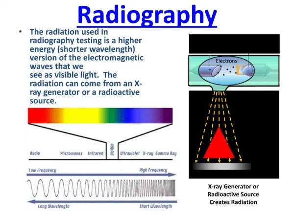
RADIOGRAPHY
Diagnostic imaging. Physical stimulus. Technique. Modality. Source. Signal. IMAGE. RADIOGRAPHY. PLANAR. X rays attenuation. X rays. DENSITY(g/cm 3 )A ATOMIC NUMBER. CT. TOMOGRAPHIC. Ultrasound Reflection (ECHO). ECHO TIME ECHO AMPLITUDE. TOMOGRAPHIC. ultrasound. ECOGRAPHY.


RADIOGRAPHY
E N D
Presentation Transcript
Diagnostic imaging Physical stimulus Technique Modality Source Signal IMAGE RADIOGRAPHY PLANAR X rays attenuation X rays • DENSITY(g/cm3)A • ATOMIC NUMBER CT TOMOGRAPHIC Ultrasound Reflection (ECHO) • ECHO TIME • ECHO AMPLITUDE TOMOGRAPHIC ultrasound ECOGRAPHY Magnetic field Radio-wave • T1 • T2 • PROTON DENSITY Magnetic resonance MR TOMOGRAPHIC PLANAR SCHINTIGRAPY Gamma rays Radiopharmaceuticals Radiation emission • RADIOACTIVITY CONCENTR. EMISSION TOMOGRAPHY SPECT e PET TOMOGRAPHIC
NUCLEAR MEDICINE ThERAPY DIAGNOSTIC IMAGING [99mTc]MDP scintigrafia planare [18F]FDG - PET [131I]NaI: scintigrafia/terapia No images without a Radiopharmaceutical except for radiation internal contaminations
I radiopharmaceutical on the basis of the EU Directive D.L.vo n.178 del 29/5/91, are classified as medicinal products. Italian version:……..è da intendersi come medicinale ogni sostanza o composizione presentata come avente proprietà curative o profilattiche delle malattie umane o animali, nonché ogni sostanza o composizione da somministrare all'uomo o all'animale allo scopo di stabilire una diagnosi medica o di ripristinare, correggere o modificare funzioni organiche dell'uomo o dell'animale.
. Aim: to carry radioactivity at target site to treat a clinical conditions (tumour) or to perform a diagnosis. A radiopharmaceutical need different kinetics and pharmacological properties depending on the use (therapy, diagnosis) or target localization (intracerebral, intracellular etc..). Afteradministration (in general by i.v. injection) the moleculereachs the target organbutalsoothers body districtsthatcouldinterefere with the quality of the images obtained or in case of therapeutic use increaseitstoxicity . Administration: intra venous
Radiofarmaceuticals are defined: Radionuclide (Radioisotope) 11 OCH3 Chemical Structure That includes the radionuclide R Radiopharmaceutical
Chemical structure: Kinetics of distribution, biological half life, target interaction.
Figure 1. Surface chelation model of 64Cu-labeled Structures • Small molecules • Peptides e antibody • Cells • Nanovectors
Radionuclides: applications Radiationemitted (type and energy), interaction with cells and tissues (radiotoxicity), halflìfe (time necessary to the 50% of atomspresent in the sample decay and are transformed in an otherelement). • Diagnostic : high penetration and low interaction with tissues, energy adequate to be detected by medical devices. Radionuclides determin the acquisition device used and the temporal frame of the potential detection of the radiopharmaceutical (half-life) • Radioisotopes gamma emitters (planar scintigraphy, or tomographic scintigraphy SPECT): 123I (10h), 99mTc (6h)… • Radioisotopes beta + or positron emitters : 11C (20.38 min.), 18F (108.9 min.), 64Cu (12.7 h.), 13N(9.97 min.) • Therapeutic: high interaction with tissue, low penetration. • beta-: 90Y, 188Re , 131I, 166Lu • Diagnostic and therapy:64Cu (beta+ and beta -), 166Lu (gamma and beta -) may be used for therapy and diagnostic
The atom Nucleus diameter 10-15m Atomic diameter 10 –10m
Elements Z Number of proton A Number of proton plus neurtrons
AX Z STRUTTURA DI UN NUCLEO 131I 53 ATOMIC NUMBER Z N° OF PROTONS MASSA NUMBER A N° OF NEUTRONS + PROTONS
Proton: positive charge particles, Kg:1,672x10-27 Neutron, kg: 1,675x10-27 Electron: negative charge particles, kg: 9,109x10-31
An element has the same number of proton An isotope of an element has the number of proton but a different number of neurtron
H 1 He 2 Proton modification: new element Helium 2 neutron +1proton H 1 H 1 H 1 Neutron: new isotop 3 1 2 Hydrogen 0 neutron Deuterium 1neutron Trizium 2 neutron + 1 neutron Electron: ionization, different reactivity
131I 127I 53 53 NUCLIDS ISOTOPS – nuclids of the same element (= Z but different n°neutrons) isotopes of a same element have similar chemical properties ISOTOPS 53 protons 78 neutrons 131 A 53 protons 74 neutrons 127 A
Radionuclides production: Reactor Cyclotron (internal use for radiolabeling or distribution) Generator (nuclear medicine radiopharmaceutical lab)
RADIOACTIV ISOTOPES PRODUCTION Radioactive may be produced by bombardament of stable nuclei with neutrons or proton that cause nuclear reaction and transformation of the stable nucleusin a radioactive unstable nucleaus REACTOR CYCLOTRONE
*Positron emitters:F-18, C-11, Zr-89, Cu-64 Gamma emitters: I-131 etc.. target *Medical cyclotrons have an energy of 11-18 MeV
Radionuclide parent Radionuclide daughter ln A t GENERATOR Tc-99m; Ga-68 The generator is a system form by: • Radionuclide “parent” long T1/2 • Radionuclid “daughter” shorter T1/2 Difference in T1/2 : almost 10 times If T1/2 of the parent isotope is > then that of daughter T1/2 after a certain time the system reach an equilibrium condition and the ratio between parent and daughter activity became constant (around unit) A t
Radiopharmaceuticals for diagnostic use: in vivo image or measure of a biologicalprocessthathas a diagnosticmeaning (or of researchinterest) • radiopharmaceutical = a tracer Ittraces a biologicicalprocesswithoutmodify the system under study. Low mass administer and high sensitive methodsrequested (able to detectsubstances in concentration < nM) • Itisnecessary a physicalsystemable to detecradioationemitted • Radiopharmaceuticals for therapy: tissuedamagespecific for a certaintype of districtthrough the use of radiationemitter by an isotope carried by a moleculeselectivefor a target preferentiallyexpresed by the target tissue (cancer) and retained in thatregion for a time sufficient to exertitstherapeuticactivity.
CT PET Diagnostic: to selectively accumulate in the target region reaching radioactivity levels a) proportional to target expression of function b) in a time adequate for the half life of the radioisotope used c) with high signal to background ratios (fast clearance from non target tissues particularly if surrounding target tissue) and with a tracer behaviour (no pharmacological activity at the target site; low target occupancy. ISOTOPES: GAMA EMITTERS: 99mTc; 111In; 123/131I Positron emitters: 68Ga, 18F, 11C, 64Cu
H CH3 OH 11C N H CH3 H RADIOPHARMACEUTICAL FOR DIAGNOSTIC USE ARE TRACERS TRACER: traces (traccia)a process. In this case biological RADIOPHARMACEUTICAL:tracer labeledwith a radioactive isotope that can be deteced by medical devices used in Nuclear Medicine A TRACER SHOULD Specific interaction with the target Mass << then the minimal mass that exert an effect Specific activity: high and adequate to detect modification in the traced signal.
Therapeutic: presence of a lesion that selectively express a certain type of target in comparison with normal tissues. The radiopharmaceutical should a) accumulate selectively in the target reagion for a time sufficiently long to affect cancer cells, b) fast kinetics from normal tissue including elimination organs (live, kidney etc.). It is not a tracer Beta minus emitters: 131I (also gamma), 90Y, 177Lu, 188Re, 64Cu (also positron) Conc. regione target regioni non target, organi eliminazione tempo
Kinetics, aims UPTAKE AT TARGET SITE RETANTION PROPORTIONAL TO TARGET EXPRESSION/FUNCTION AND NOT OTHER VARIABLES CLEARANCE: FAST CLEARANCE FROM NON TARGET REGIONS LACK OF RADIOACTIVE METABOLITES AND IF PRESENT NOT AVAILABLE IN THE TARGET TISSUE
In vivo kinetics: in case of radiopharmaceuticals we can measured the kinetics of uptake and clearence at organ levels
A B KINETICS CA/CB retention Conc. A:target region Signal to noise ratio B: non target region Temporal window for measurement clearance uptake time Diagnostic/therapy [111In]-ibritumomab tiuxetan, imaging to verify the presence of CD20 in lymphoma patients using [90Y]-ibritumomab tiuxetan Acquisition: 24 hrs post injection Diagnostic: [99mTc]MIBI- Myocardial perfusion Acquisition: 1 hrs post injection
Upatake Arival of the radiopharmaceutical in the target tissue. It is the major event in the first phase of distribution. It depends on: • Organ Perfusion • Diffusion across endotelial vascular membranes and eventually across BBB or cell membrane if the target is intracellular • Plasma Protein binding or blood cell uptake
PERFUSION:delivery from blood Perfusion is the passage of fluid through the circulatory system or lymphatic system to an organ or a tissue, usually referring to the delivery of blood to a capillary bed in tissue per unit of time. It influence the early phase of distribution. It can be the only phenomenon that can be measure if it is prevalent on the other (delivery dependent radiopharmaceuticals) or if it is the biological function of interest (radiopharmaceutica for lblood flow measurement). Perfusion influences also radioactivity clearance. The higher perfusion the higher is the clearance from a certain tissue. . Conc Brain perfusion: [Tc-99m]ECD time
DIFFUSION • Intravascular target: blood volume measurement (es. radiolabeled albumine). No membranes • Extravascular but extracellular target: endotelium membrane: membrane receptor or uptake sites of peripheral organs) • Intracellular target: cell membrane (enzymes, intracellular recptors ets) 4) Brain target (BBB crossing) sangue cellula
DIFFUSION • It depends on: • Molecular ray • Proprietà chimChimico-physical properties: • pKa (ionizzation, polarity) • Liposolubily • Conc. Gradient (if passive diffision) • Presence of transporte (es: glucose and FDG, amino-acids and FET) • Protein binding or blood cell uptake
PASSIVE DIFFUSION FICK LOW Passive diffusion acros a membran depends on concentration gradient between plasma and tissue of the diffusable form (not bound, not ionized). First order kinetic process. It depends on Permeability (A,H,K,D) and concentration gradient. At the beginning the rate will be blood versus organ All’ inizio sara’ favorito passaggio sangue tessuto successivamente sara’ tessuto sangue
MOLECULAR RAY Ray (A) EQ. Diffusibility MW Coef.MO
Ionization: • POLAR COMPOUNDS (R-COOH, RNH2, gruppi –OH, -SH, -NH2). • Depends on pKa of the tracer and pH of tissues (R-COOH, RNH2 ): stomach (pH acid) –intestin (pH basic). • The higher is ionization the lower is diffusion across membrane and BBB • Liposolubility: • Non polar compounds; depends on chemical structure (aromatic groups, long C chanis, apolar roups). • Liposolubile compound, if ionic ray is adequate can be used for brain studies R-COOH R-COO- + H+
Trasporter (FDG, FET, FLT, FDOPA etc...) • Glucose (GLUT1,3 etc): [18F]FDG • Aminoacid transporter: [11C]Methionine, [18F]fluoro-ethyl-tyrosine, [18F]fluoro -tymidine • Choline tranporter: [11C]Choline
RADIOPHARMACEUTICAL for the measurement of perfusion • Aim: to measuretissueperfusio. Radioactivitydistributionshould be proportionalat a certain time afterinjection to regionaltissueperfusion. Twoclasseshavebeendeveloped • 1) Inerthighlydiffusible RF (133Xe, [15O] • H2O). Radioactivityuptake and clearance istotalperfusiondependent. • 2) Chemicalmicrospheres ([13N]NH3, [99mTc]MIBI; highlydiffusible. Whenetaken up by tissue are immediatelytransformed by highlyexpressed targets and remaintrappedinto the tissue. Blood activityisalsorapidelymetabolized and cleared so thatincrease in tissueconcentrationisnotpossible. Theirregionalconcentrationisproportional to regionalperfusion. High perfusion Tissue region Low perfusion Tissue region Blood time
TARGETS OR PROCESSES • Receptors • Enzymes • Transporter • Antigens • Reporter gene • Proteins or cells • Hypoxia hypoxia Microglia activation glycolisis
Retention This phase, describes radiopharmaceutical-target interaction. When the radiopharmaceiutical interacts with the target, the rate of clearance is lower and radioactivity is retained in tissue with a concentration that is proportional to target levels/activity. This time frame represent the diagnostic window for image acquisition in case of diagnostica agents. RF-target describes the mechanism of action of the RF. For therapeutic agents RF-taregt interaction in necessary to be retained in the target tissue and exert its radiotoxicity Diagnostic window: time frame where radioactivity concentration is proportional to target expressio and not to radioactivity delivery (perfusion etc..) Therapeutic window: time frame where RF exert its radioatocity at taregt site retention Conc. Target uptake Target interaction No target clearance
Retention M B T 1) Blood conc. > tissue conc, from B to T 2) Transient equilibrium 3) If radiotracer is not bound or trappedinto the tissue blood conc drop and direction is T to B Uptake phase Steady state Clearance phase B=blood (art or ven) T=tissue M=membranes
RETENTION • Biotransformation and trapping • Binding
Biotransformation and trapping • The radiopharmaceutical is transformed in a different chemical entity but manteining the radionuclide in its structure in general by an enzimatic reaction. The new chemical entity has a clearance rate from tissue >> slower than that of the original radiopharmaceutical or is trapped by tissue (clearance = 0) during measurement time frame. Tissue radioactivity increase with time reaching a maximum uptake that is proportional to the amount and activity of the target. The product is less soluble, bound to some intracellular component etc.. • Glycolisis: FDG FDG-6P (FDG=fluorodeoxyglucose) • Cell proliferation: FLT FLT 6P (FLT=fluoro-L-tymidine)
BBB tissue plasma Cp F-18]FDG F-18]FDG-P [F-18]FDG GLUT HK [18F]fluorodeoxyglucose No to* Fructose -6-P Conc FDG+FDG6-P *Absence of the hydroxylgroup in 2 precludes the isomerization from an aldehyde (glucose) to a ketone (fructose) chain and remeintrapped inside the cell 60 min. FDG Time (min)
Target region non target region Binding Specific binding between RF and receptors, enzymes, transporter. Binding is reversible and the traget is saturable. RF+target [RF-Target] RF-target binding reduces the rate of clearence of radioactivity fro tissue.
Binding • Recettor or transporters: • [11C]Raclopride- DA-D2like rec • [123] DATscan Dopamine transporter. • [68Ga]DOTATOC or DOTATATE. • [68Ga]PSMA • Proteins • [11C]PIB – beta amyloid • New radiopharmaceuticals for tau
For a new Radiopharmacology it is crucial : • Quality of preparation or production • Efficacy: optimal acquisition, absence of radioactive metabolites at target site, time or therapeutic scheme (therapy) • Safety: radioexposure and the absorbed dose (dosimetry). c
40 – 80 MIN 0 – 10 MIN 5HT2a SEROTONIN REC. ARE PRESENT IN CORTECCIA (CTX) BUT NOT IN THE CEREBELLUM( CER). Cerebellum uptake at early time depends on tissue perfusion and RF delivery RETENTION EARLY UPTAKE Ctx MBq/ml Cer 90 min.
A B total-body images obtained with a gamma camera immediately after the injection of l (A) and 24 hrs. After (B) [111In]-ibritumomab tiuxetan an antibody for CD20. This imaging exam is preliminary to the administration of the therapeutic RF 90Y-ibritumomab tiuxetan (that bindind an is taken up by lymphoma cells CD20 positive). At 24 hrs but not a 1 hr. it possible to detect radioactivity uptake at the level of a lymphonode. Diagnostic window: 24 hrs.