Download

1 / 16
200 likes | 574 Vues
Digital radiography and conventional analogue radiography. Narufumi Suganuma Division of Environmental Health Department of International Social and Health Sciences University of Fukui School of Medicine. Instruction. Read the film numbered #1 to 16 on the each view box.

E N D
Digital radiography and conventional analogue radiography Narufumi Suganuma Division of Environmental Health Department of International Social and Health Sciences University of Fukui School of Medicine
Instruction • Read the film numbered #1 to 16 on the each view box. • Finish reading each film in 3 min and move to the next desk to the right. • Continue the procedure until you finish reading all the 16 films.
Standard films • Full set • 0/0, 0/0 • 1/1p/p, 2/2p/p, 3/3p/p • 1/1q/q, 2/2q/q, 3/3q/q • 1/1r/r, 2/2r/r, 3/3r/r • 1/1s/t, 2/2s/s, 3/3s/s • 1/1t/t, 2/2t/t, 3/3t/t • u/u quad • A, B, C • Pleural abnormalities
Standard films • Full set • 0/0, 0/0 • 1/1p/p, 2/2p/p, 3/3p/p • 1/1q/q, 2/2q/q, 3/3q/q • 1/1r/r, 2/2r/r, 3/3r/r • 1/1s/t, 2/2s/s, 3/3s/s • 1/1t/t, 2/2t/t, 3/3t/t • u/u quad • A, B, C • Pleural abnormalities
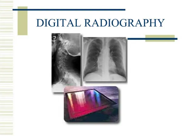
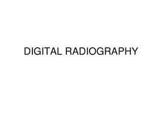
radiograph and digital technique • Analogue radiograph: film-screen radiograph • Computed radiograph: imaging plate of storage phosphor, need plate reader to obtain digital data • Digital radiograph: flat-panel detector catches X-ray, turns it to photon and obtains digital data
Introduction • Aimed to compare film quality and small opacity detection • Comparison using high-kilovoltage technique between • conventional film-screen radiography • amorphous silicon flat-panel detector (FPD) system (CXDI-11, Canon, Tokyo)
Trial materials were: • AR, DR, DRe, DRc • 1, 6, 11, 16 = normal control 0/0 • 2, 7, 12, 14 = early asbestosis 0/1,1/0 • 3, 5, 10, 15 = silicosis 1/1 • 4, 9 = severe silicosis 3/2 • 8, 13 = severe asbestosis
Subjects and methods • Eleven physicians have independently evaluated film quality and classified profusion of 11 pairs of conventional radiograph and film taken by FPD with one week’s interval. • Film quality was evaluated with ILO film quality assessment grade with 4-point scale, considering technical defects concerning density, contrast, positioning and so on.
Results 1 • Using 4-point scale film quality (excellent, good, fair or acceptable), FPDs’ quality fell in excellent or good, whereas conventional radiographs’ quality varied from good to acceptable. • Mode of 11 physicians’ reading results was consistent between conventional radiograph and FPDs. • These results were also consistent with CT assisted reading results of another radiologist.
Results 2 • Cases with considerable inconsistency of reading results were those with higher profusion p-type opacities, and a case with emphysema.
CXR Case 8 FPD
Case 8 FPD CXR
CXR Case 11 FPD
Case 11 FPD CXR
Conclusion • FPDs showed constantly better film quality than conventional radiograph. • ILO classification results of small opacities profusions were not largely different between conventional radiograph and FPD. Acknowledgement We thank technical support for film preparation to Mr. Takashi Ogura, Canon.