Download
1 / 1
10 likes | 101 Vues
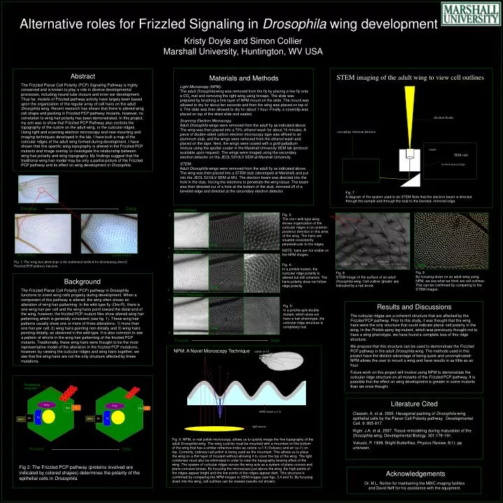
Alternative roles for Frizzled Signaling in Drosophila wing development. Fritz. Fritz. Fy. In. Fy. In. Proximal. Distal. Kristy Doyle and Simon Collier Marshall University, Huntington, WV USA. Pk. Stbm. NPM. Wild Type Wing. prickle mutant. Abstract

E N D
Alternative roles for Frizzled Signaling in Drosophila wing development Fritz Fritz Fy In Fy In Proximal Distal Kristy Doyle and Simon CollierMarshall University, Huntington, WV USA Pk Stbm NPM Wild Type Wing prickle mutant Abstract The Frizzled Planar Cell Polarity (PCP) Signaling Pathway is highly conserved and is known to play a role in diverse developmental processes, including neural tube closure and inner ear development. Thus far, models of Frizzled pathway activity have largely been based upon the organization of the regular array of cell hairs on the adult Drosophila wing. Recent research has shown that there is altered wing cell shape and packing in Frizzled PCP pathway mutants, however, no correlation to wing hair polarity has been demonstrated. In this project, my aim was to show that Frizzled PCP Pathway also controls the topography of the cuticle on the adult wing, or the cuticular ridges. Using light and scanning electron microscopy and new mounting and imaging techniques developed in the lab, I have built a map of the cuticular ridges of the adult wing formed during development. I have shown that this specific wing topography is altered in the Frizzled PCP mutants and image overlay to investigate the relationship between wing hair polarity and wing topography. My findings suggest that the traditional wing hair model may be only a partial picture of the Frizzled PCP pathway and its effect on wing development in Drosophila. STEM imaging of the adult wing to view cell outlines Materials and Methods Light Microscopy (NPM):The adult Drosophila wing was removed from the fly by placing a live fly onto a CO2 mat and removing the right wing using forceps. The slide was prepared by brushing a fine layer of NPM mount on the slide. The mount was allowed to dry for about ten seconds and then the wing was placed on top of it. The slide was then allowed to dry for about 1 hour. Finally, a coverslip was placed on top of the dried slide and sealed. Scanning Electron Microscopy:Adult Drosophila wings were removed from the adult fly as indicated above. The wing was then placed into a 70% ethanol wash for about 10 minutes. A piece of double-sided carbon electron microscopy tape was affixed to an aluminum stub, and the wings were removed from the ethanol wash and placed on the tape. Next, the wings were coated with a gold-palladium mixture using the sputter-coater in the Marshall University SEM lab (protocol available upon request). The wings were imaged using the secondary electron detector on the JEOL 5310LV SEM at Marshall University. STEM: Adult Drosophila wings were removed from the adult fly as indicated above. The wing was then placed into a STEM stub (developed at Marshall) and put into the JEOL 5310LV SEM at MU. The electron beam was directed into the hole in the stub, forcing the electrons to penetrate the wing tissue. The beam was then directed out of a hole at the bottom of the stub, mirrored off of a beveled edge and directed at the secondary electron detector. electron beam secondary electron detector sample TEM stub Fig. 8STEM image of the surface of an adultDrosophila wing. Cell outline ‘ghosts’ are indicated by a red arrow. beveled mirrored edge Fig. 7A diagram of the system used to do STEM Note that the electron beam is directed through the sample and through the stub to the beveled, mirrored edge. pkpk-30 homozygote A P Fig. 3: The ore-r wild type wing shows organization of the cuticular ridges in an anterior-posterior direction in this area of the wing. The hairs are situated consistently perpendicular to the ridges. NOTE: hairs are not visible on the NPM images. Oregon R - wild-type A P ore-R (wild type) Fig. 4: In a prickle mutant, the cuticular ridge polarity is altered but still coherent. The hairs polarity does not follow ridge polarity. NPM Fig. 9By focusing down on an adult wing using NPM, we see what we think are cell outlines. This can be confirmed by comparing to the STEM images. Background The Frizzled Planar Cell Polarity (PCP) pathway in Drosophila functions to orient wing cells properly during development. When a component of this pathway is altered, the wing often shows an alteration of wing hair patterning. In the wild-type fly (Ore-R), there is one wing hair per cell and the wing hairs point toward the distal end of the wing, however, the frizzled PCP mutant flies show altered wing hair patterning which is generally consistent (see fig. 1). These wing hair patterns usually show one or more of three alterations: 1) more than one hair per cell; 2) wing hairs pointing non-distally and 3) wing hairs pointing distally, as observed in the wild type. It is also common to see a pattern of whorls in the wing hair patterning of the frizzled PCP mutants. Traditionally, these wing hairs were thought to be the most representative model of the alteration in the frizzled PCP mutations, however, by viewing the cuticular ridges and wing hairs together, we see that the wing hairs are not the only structure affected by these mutations. NPM A P Fig. 5:In a prickle-sple double mutant, which does not have a hair phenotype, the cuticular ridge structure is completely lost. pkpk-sple14 homozygote Results and Discussions The cuticular ridges are a coherent structure that are affected by the Frizzled PCP pathway. Prior to this study, it was thought that the wing hairs were the only structure that could indicate planar cell polarity in the wing. In the Prickle-spiny leg mutant, which was previously thought not to have a wing phenotype, we have found a complete loss of this ridge structure. We propose that this structure can be used to demonstrate the Frizzled PCP pathway in the adult Drosophila wing. The methods used in this project have the distinct advantage of being quick and uncomplicated: NPM allows the user to mount a wing and have results in as little as an hour. Future work on this project will involve using NPM to demonstrate the cuticular ridge structure on all mutants of the Frizzled PCP pathway. It is possible that the effect on wing development is greater in some mutants than we once thought. Fig. 1: The wing hair phenotype is the traditional method for determining altered Frizzled PCP pathway function. NPM Proximal Distal NPM: A Novel Microscopy Technique cuticle =1.5 Developing wing hair Dsh Fz Dsh Fz Pk Stbm NPM mount =1.5 Literature Cited light source Classen, A. et al. 2005. Hexagonal packing of Drosophila wing epithelial cells by the Planar Cell Polarity pathway. Developmental Cell. 9: 805-817. Kiger, J.A. et al. 2007. Tissue remodeling during maturation of the Drosophila wing. Developmental Biology. 301:178-191. Vukusic, P. 1998. Bright Butterflies. Physics Review. 8(1): pp unknown. Fig. 6: NPM, or nail polish microscopy, allows us to quickly image the fine topography of the adult Drosophila wing. The wing (cuticle) must be mounted with a mountant on the bottom of the wing that has a similar refractive index as cuticle =1.5 (Vukusic) and air (=1) on top. Currently, ordinary nail polish is being used as the mountant. This allows us to place the wing on a thin layer of moutant without allowing it to cover the top of the wing. The light condenser must also be eliminated in order to view the topography lensing effect of the wing. The system of cuticular ridges across the wing acts as a system of plano-convex and plano-concave lenses. By focusing the microscope just above the wing, the high points of the ridges appear bright and the low points of the ridges appear dark. This structure is confirmed by comparing the NPM images to SEM images (see figs. 3,4 and 5). By focusing down into the wing, cell outlines can be viewed (results not shown). Proximal Distal Fig 2: The Frizzled PCP pathway (proteins involved are indicated by colored shapes) determines the polarity of the epithelial cells in Drosophila. Acknowledgements Dr. M.L. Norton for maintaining the MBIC imaging facilties and David Neff for his assistance with the equipment.