Download

1 / 11
110 likes | 252 Vues
This document delves into crucial anatomical and pathological aspects of synovial joints and bone conditions, as demonstrated through questions from a 2004 past exam. It emphasizes the distinctive features of synovial joints, highlighting the joint capsule, synovial membrane, and fluids. The discussion also involves evaluating radiographs in trauma cases, testing structures like the popliteal artery and nerves, and addressing common errors in interpreting bone disorders. Additionally, it covers common bone tumors, signs of osteomyelitis, and risk factors associated with different age groups. ###

E N D
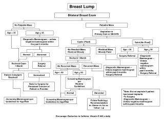
A lump in the leg Quiz
2004 past exam Q • Name the characteristic features of a synovial joint • Joint capsule • Synovial membrane • Synovial fluid / joint cavity • Articular cartilage • Bone
2004 past exam Q This is a radiograph of the knee of a patient who has been involved in a motor vehicle accident. List three (3) structures whose integrity you must test prior to treating this injury. • Popliteal artery • Tibial nerve • Peroneal nerve • ?? others
2004 past exam Q’s – TRUE or FALSE Osteogenesisimperfecta is a disorder of defective synthesis of collagen type I. True Metastases in bone usually cause a periosteal reaction. False Most osteosarcomas occur before the age of thirty (30) years. True Osteosarcomas are aggressive tumours. True Sclerotic bone around the edge of a lesion in a bone, is a poor prognostic sign. False Giant cell tumours (osteoclastoma) are usually benign. True
Feedback:- November 2004 Many students confused film exposure with osteoporosis Confusion of the growth plates and sutures as fractures Many stated that the x-ray was of degenerative changes despite being from a young child ie osteoporosis and osteoarthritis giving fractures. (How many could tell this was a child’s x-ray?) Confusion of fovea capitis with an abnormality Confusion of ileum and ilium Confusion of hip joint with sacroilac joint Some students couldn’t tell left from right despite it being written on the radiograph. One student claimed a diagnosis of hepatitis, although osteoporosis and fractures and malignancies were commonest. Common Errors:- Lack of knowledge in interpreting radiographs. The majority didn’t realise it was a child’s x-ray or didn’t actually notice the growth plate lucencies, then they decided that it was osteoarthritis/osteoporosis and then went looking for evidence to support their ideas.
Knee examination – Mix & Match • Collateral ligaments • Cruciate ligaments • Meniscuses
Osteochondroma Enchondroma Simple bone cyst Osteosarcoma Osteoidosteoma Chondroblastoma Giant cell tumor Ewing’s tumor E A B F C G H D
Tumours of bone and cartilage • Malignant, 15-30yo, around the knee, sunburst periosteal reaction • Usually benign. Giant multinucleated cells. • Small sclerotic central nidus • Benign cartilage cyst in bone marrow • Small round blue cells • Rare, malignant, notochord remnant • Cartilage-capped exostosis (bony outgrowth) Osteosarcoma Giant cell tumour Osteoidosteoma Enchondroma Ewing’s sarcoma Chordoma Osteochondroma
Osteomyelitis A patient presents with dull unilateral hip pain, progressing over a week. If this is a case of osteomyelitis, what local and systemic signs and symptoms might you expect? Tenderness, warmth, erythema, swelling, fever and rigor may also be present. However, patients with osteomyelitis involving sites such as the hip, vertebrae, or pelvis tend to manifest few signs or symptoms other than pain
Osteomyelitis The risk of osteomyeletis spreading from the bone shaft to the bone end and the into a joint depends on age. Considering infants, children, and adults, which groups are at higher risk and why? Infants have capillaries bridging the growth plate. Adults have the growth plate resorbed.