Download
1 / 62
660 likes | 1.13k Vues
NEUROANATOMY Lecture : 7 Blood Supply of the Brain, Veins of the Brain & The Cerebral Dural Sinuses. Prepared and presented by: Dr. Iyad Mousa Hussein, MD, Ph.D in Neurology Head of Neurology Department Nasser Hospital. LECTURE OBJECTIVES:.

E N D
NEUROANATOMY Lecture : 7 Blood Supply of the Brain, Veins of the Brain & The Cerebral Dural Sinuses Prepared and presented by: Dr. Iyad Mousa Hussein, MD, Ph.D in Neurology Head of Neurology Department Nasser Hospital
LECTURE OBJECTIVES: Origin, course & Branches of the Internal Carotid Arteries. Origin, Branches, and Clinical importance of the Anterior Cerebral Arteries. Origin, Branches, and Clinical importance of the Middle Cerebral Arteries. Origin, course & Branches of the Vertebral Arteries. Origin, Termination & Branches of the Basilar Artery. Origin, Branches, and Clinical importance of the Posterior Cerebral Arteries. Circle Of Willis Formation. The important anastomoses between the cerebral arteries. Veins of the Brain. The Cerebral Dural Venous Sinuses.
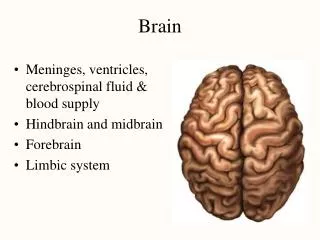
Blood Supply of the Brain • Brain blood supply formed of two main system (arteries): • Carotid system: Right & Left internal carotid artery (ICA). • 2.Vertebrobasilar system: Right & Left vertebral artery (VA). • Normal blood flow for the entire brain is 750 ml/min.
Branches of the External Carotid Artery 1. Superior thyroid artery. 2. Ascending pharyngeal artery. 3. Lingual artery. 4. Facial artery. 5. Occipital artery. 6. Posterior auricular artery The ECA divided into two terminal branches: a. Superficial temporal artery. b. Maxillary artery.
I. The Internal Carotid Artery Origin: arises in the neck of the terminal branches (at bifurcation) of the common carotid artery (CCA). Each ICA enters cranial cavity through carotid canal. Course: The ICA has 4 parts: 1. Cervical part. 2. Intrapetrous part. 3. Intracavernous part. 4. Intracranial part.
Branches of the Internal Carotid Artery 1. Ophthalmic Artery: which enters the optic canal below the optic nerve to supply the orbit & gives central retinal artery. 2. Posterior Communicating Artery: which joints the posterior cerebral arteries to establish the circle of Willis. 3. Anterior Choroidal Artery. 4. Anterior Cerebral Artery. 5. Middle Cerebral Artery: the larger branch & the main trunk of the ICA.
Anterior Choroidal Artery Origin: from the ICA just before it divides into its two terminal branches (anterior & middle cerebral arteries). It supplies: the optic tract, lateral geniculate body (LGB), internal capsule and choroid plexus of inferior horn of lateral ventricle.
Anterior Cerebral Artery (ACA) Origin: it arises from the ICA. It becomes joined to other anterior cerebral artery of the opposite side by anterior communicating artery.
Branches of the Anterior Cerebral Artery • Cortical Branches: to supply the medial surface of the hemisphere from frontal pole to the parieto-occipital sulcus. • 2. Central Branches: to supply the anterior part of the corpus striatum, the anterior part of anterior limb internal capsule & the septal region. • 3. Callosal Branches: to supply corpus callosum.
Clinical importance of Anterior Cerebral Artery The anterior cerebral artery supplies three important regions: The motor & sensory areas of the lower limb in the precentral gyrus. The corpus callosum: obliteration of its blood supply may result in apraxia.
Middle Cerebral Artery (MCA) Origin: it arises from ICA as the larger of the two terminal branches.
Branches of the Middle Cerebral Artery • Cortical Branches: to supply lateral surface of the cerebral hemisphere, the lateral half of the orbital surface of the hemisphere, the temporal pole. • 2. Central Branches: are many branches called striate arterioles to supply the corpus striatum (lentiform & caudate) and internal capsule. • One of the central branches is larger than the others & is • called the artery of cerebral hemorrhage (Chorcot's • artery).
Clinical Importance of Middle Cerebral Artery It supplies: 1. The motor and sensory areas for the whole body except the lower limbs. 2. Most of the internal capsule, obstruction of its blood supply → hemiplegia. 3. The auditory area in the superior temporal gyrus. 4. Motor speech area in the inferior frontal area → expressive aphasia.
II. The Vertebrobasilar System • Vertebral Artery • Origin: each vertebral artery arises from the first part of subclavian artery. • Course: • From subclavian artery → then passes upwards • through the transverse foramina of the upper 6 cervical vertebrae → foramen magnum → at the lower border of pons two vertebral arteries unite to form the single Basilar Artery.
Branches of Vertebral Artery 1. Posterior Inferior Cerebellar Artery. 2. Posterior Spinal Artery. 3. Anterior Spinal Artery. 4. Medullary Branches.
Basilar Artery Origin: at the lower border of pons by the union of the two vertebral arteries. It ascends along the basilar sulcus infront of the pons. Termination: at the upper border of the pons by dividing into two posterior cerebral arteries.
Branches of Basilar artery 1. Pontine (Paramedian) Branches. 2. Anterior Inferior Cerebellar Artery. 3. Internal Auditory (Labyrinthine) Artery. 4. Superior Cerebellar Artery.
Posterior Cerebral Artery (PCA) Origin: the right & left posterior arteries arise at the upper border of the pons as the two terminal branches of the basilar artery.
Branches of Posterior Cerebral Arteries 1. Cortical Branches: to supply occipital lobe and inferior border of temporal lobe. 2. Central Branches (Short Medial and Long Lateral): to supply anterior part of thalamus, mammilary bodies, subthalamic region and lateral side of midbrain. 3. Posterior Choroidal Arteries: to supply the choroid plexus of the lateral and third ventricle.
Clinical importance of Posterior Cerebral Arteries It supplies: 1. The whole visual cortex in the occipital lobe. 2. Most of the midbrain. 3. Most of the thalamus. 4. The centre of smell in the uncus 5. Most of the choroid plexus of the lateral and third ventricles.
Circle Of Willis Definition: it is an arterial anastomoses between Nine arteries supplying the brain. Function: Direct blood to other branches in cases of occlusion and connect carotid system with vertebrobasilar system.
Circle Of Willis Formation 1. The right & left of anterior cerebral arteries. 2. The anterior communicating artery connecting the two anterior cerebral arteries. 3. The right & left internal carotid arteries. 4. The right & left posterior communicating arteries connecting the posterior cerebral arteries with internal carotid arteries. 5. The right & left posterior cerebral arteries.