EPR Signal Calibration and Photosynthesis Response in Chlamydomonas reinhardtii Under Varying Conditions
This study investigates the correlation of EPR signals based on H2O2 concentration, using a calibration curve established with H2O2-derived hydroxyl radicals. The experiment involved incubating specific concentrations of H2O2 in TAP medium along with 4-POBN, ethanol, and Fe-EDTA to assess radical formation. In parallel, the photosynthetic response of wild-type Chlamydomonas reinhardtii and catalase knock-down mutants was assessed following a shift from darkness to high light. Results illustrate the impact of catalase activity on photosynthesis under varying light conditions.


EPR Signal Calibration and Photosynthesis Response in Chlamydomonas reinhardtii Under Varying Conditions
E N D
Presentation Transcript
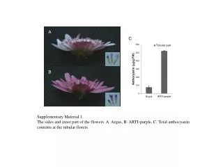
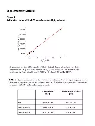
Supplementary Material Figure 1 Calibration curve of the EPR signal using an H2O2 solution Dependence of the EPR signals of H2O2-derived hydroxylradicals on H2O2 concentration. A given concentration of H2O2wasadded to TAP medium and incubated for 5 min with50 mM 4-POBN, 4% ethanol, 50 μM Fe-EDTA. Table 1: H2O2 concentration in the cultures as determined by the spin trappingassay. Chlorophyll concentration of the culture: 10 µg ml-1. Results are expressed as mean bars represent ± S.D. (3-6 independent experiments).
Figure 2 Photosynthesis after a dark to high light switch in wild type C. reinhardtii and in catalase knock-down mutants. wild type amiRNAcat40 amiRNAcat15 Photosynthesis was measured in the presence (circles) or absence (diamonds) of 1 mMNaHCO3 in wild type and catalase knock-down mutants exhibiting 40% or 15% residual catalase activity following a dark to high light (700 µmol quanta m-2s-1) shift, and during an additional 30 min recovery at 70 µmol quanta m-2s-1. Typical O2 evolution curves are shown, representative of at least three independent experiments. Measurements of photosynthesis were performed in a Liquid-Phase Oxygen Electrode Chamber (Hansatech Instruments, Norfolk, England). Photosynthetic activity was measured in TAP medium under saturating white light.