Download
1 / 44
540 likes | 1.88k Vues
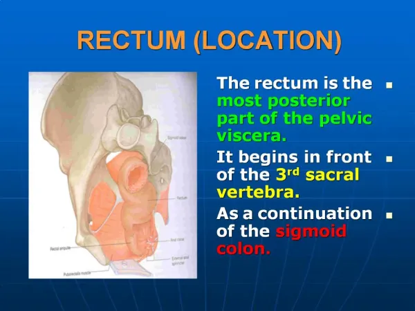
Diseases of Rectum and Anal Canal. Anatomy I. Rectum – distal part of gastrointestinal tract It´s about 14 – 16 cm long and it´s divided into three parts: 1. proximal part 2. middle part 3. distal part ( anal canal ). Anatomy II.

E N D
Anatomy I. Rectum – distal part of gastrointestinal tract It´s about 14 – 16 cm long and it´s divided into three parts: 1. proximal part 2. middle part 3. distal part ( anal canal )
Anatomy II. Blood supply : 1. superior rectal artery ( inferior mesenteric artery ) 2. two middle rectal arteries ( internal iliac artery ) 3. two inferior rectal arteries ( internal pudendal artery ) Internal rectal venous plexus - lies in the submucosa of the anal canal above the level of the dentate line ( internal haemorrhoids) External rectal venous plexus - lies under the skin of the anal canal below the dentate line ( external haemorrhoids )
Congenital abnormalities I. Imperforate anus – one infant in 4500-5000 is born with imperforate anus A. Low abnormalities : anal stenosis ( dilatation ) anal membrane - anus is covered with a thin membrane ( incision ) B. High abnormalities : ano – rectal agenesis ( 80-85 % ), often with recto-urethral or recto - vaginal fistula rectal atresia – anal canal is normal but ends blindly above the pelvic floor
Congenital abnormalities II. Examination : inspection, X-ray picture ( infant is held upside down with the coin or metal button in the site of the anus and the gas in the rectum will rise to the top and indicate the distance ) Treatment : operation incisio, dilatation, colostomy, reconstruction of the anorectum
Fissura-in-ano I. - longitudinal ulcer in the distal part of anal canal The site of location: - mid-line posteriorly - 80% - mid-line anteriorly - 10% - lateral – 10 % ( Crohn´s disease ) Ethiology – unknown ( passage of a hard stool ) - resting anal pressure is raised, but this may be due to secondary sphincter spasm induced by pain Two types: 1. acute 2. chronic ( hypertrophic anal papila and sentinel tag )
Fissura-in-ano II. Symptoms : pain, bleeding, pruritus,constipation.discharge Management : 1. conservative ( acute) - sitz baths, laxatives, anal dilatation, local creams 2. operation ( chronic) - excision of fissura, posterior or lateral sphincterotomy to reduce the high resting anal pressure
Haemorrhoids ( Piles ) I. Haemorhoids ( the dilatate rectal venous plexus ) consist of an internal and external component ( haemorhoideal disease ). - very frequent disease Ethiology - hereditary ( weakness of the vein walls ) - higher pelvic pressure ( pregnancy ), - constipation, straining at stool
Haemorrhoids II. Symptoms : bleeding, prolapse of nodes, pruritus, pain, discharge Diagnosis: inspection - at 3,7 and 11 o´clock in litothomy position rectoscopy, anoscopy Complications : bleeding, thrombosis, inflamation
Haemorrhoids III. Classification: 4 degrees I. degree : occasional bleeding II. degree : prolapse after defecation with spontaneous reposition III. degree : prolapse nodes need to be replaced manually IV. degree : permanet prolaps with inflamation, thrombosis etc.
Haemorrhoids IV. Management : A. conservative : sitz baths, local creams and suppositories, venotonics B. semoconservative : injection sclerotherapy, infrared coagulation, rubber band ligation C. operative treatment : - open and closed haemorhoidectomy - PPH procedure ( no external skin wound, recovery is rapid and relatively pain free )
Anal Abscess and Fistula I. Anal abscess and fistula are two phases of the same disease. Abscess - acute phase Fistula – chronic phase Ethiology : - majority of abscesses originate in the intersphincteric space from infection of anal gland.
Anal Abscess and Fistula II. Fistula-in-ano ( anal fistula ) usually consists of : - internal opening - primary tract - external opening Primary tract connects the internal and external openings.
Anal Abscess and Fistula III. Symptoms : acute abscess – pain, fewer fistula- in- ano – chronic purulent discharge Management : Acute abscess – surgical inicision and drainage cavity is dressed with gause ( changing every 24 hours ) wound is left open for secondary healing Anal fistula – treatment according to the type of fistula 1. discision ( lay open the primary track ) 2. drainage cum seton 3. advancement flap
Rectal Tumors 1 . Benign tumors 2. Malignant tumors
Benign rectal tumors The most frequent are polyps. Polyp is a localised elevated lesion arising from an epithelial surface. Polyp - adenoma : 90% - other ( inflammatory, hyperplastic etc. ) : 10% 2 types of adenoma : tubular ( pedunculated ) 20% villous ( sessile ) 80% Symptoms : bleeding, producing of mucus ( villous ) Treatment : polypectomy by colonoscopy surgical excision – large sessile polyp
Rectal carcinoma I. Rectal cancer ( adenocarcinoma ) arising from the epithelial cells of the rectal mucosa. 50% of all colorectal tumors are located in the rectum. Prognosis is related to typing, grading and staging. Typing : adenocarcinoma signet ring cell carinoma ( with producing of mucus ) , melanoma Grading : - well differentiated carcinoma - moderate - poor Staging : Dukes classification (ABC), TNM classification
Rectal carcinoma II. Symptoms: bleeding, sense of incomplete defecation, alteration in the bowel habit, passage of mucus Diagnosis: rectal examination , rectoscopy, colonoscopy,biopsy, barium enema examination, endorectal ultrasonography, abdominal USG, CT Dissemination: local spread, lymphatic spread, venous spread
Rectal carcinoma II. Treatment : - surgical terapy ( radical OP, paliative OP) - combined therapy ( surgery and oncotherapy ) Radical operations: anterior resection, abdominoperineal resection, radical excision Paliative operations : cryotherapy, colostomy, stents - classic or laparoscopic procedures, T.E.M.