Download
1 / 49
620 likes | 1.42k Vues
Anesthetic Considerations In Patients with Cardiac Pacemaker. DR . RAHUL NORAWAT. University College of Medical Sciences & GTB Hospital, Delhi. CONTENTS. History. Battery-operated pacing devices was introduced by C. W. Lillehei and Earl Bakken in 1958.

E N D
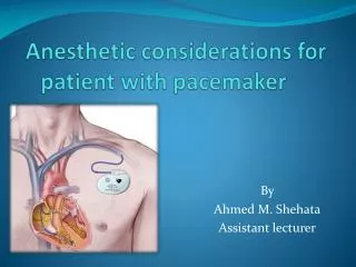
Anesthetic Considerations In Patients with Cardiac Pacemaker.. DR. RAHUL NORAWAT University College of Medical Sciences & GTB Hospital, Delhi
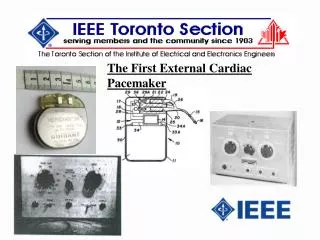
History • Battery-operated pacing devices was introduced by C. W. Lilleheiand Earl Bakkenin 1958. • In 1960, Wilson Greatbatch, created the first implantable battery-powered device. • Invention of the implantable cardioverter-defibrillator (ICD) in 1980 by Michael Morchower. First approved by U.S. Food and Drug Administration (FDA) in 1985.
INTRODUCTION • 30,00,000 patients worldwide have been implanted pacemakers while 3,00,000-5,00,000 have a Implantable Cardioverter Defibrillator (lCD). • 1,15,000 new devices are implanted each year in U.S. Rozner MA. Anaestheslol. 2007;20:261-268.
Temporary Pacing Transcutaneous Pacing • External electrode pads and power device • Large electrodes over precordium & back at level of heart • Output: 0-140 mA • Terminate: Tachy-arrhythmia • Pacing rate: 0-180 bpm • Sensing facility (VVI pacing possible) Transvenous Pacing • Electrode placed via femoral, brachial, IJV or subclavian vein • 90% success rate in absence of fluroscopy under ECG guidance • Atrial J-shaped electrodes and balloon tipped ventricular electrodes • Externally paced generator with output 0-l0mA • All pacing modes available
Temporary Pacing Indications • Unstablebradydysrhythmias • Unstabletachydysrhythmias • Third degree Atrioventricular block • Endpoint reached after resolution of the problem or permanent pacemaker implantation.
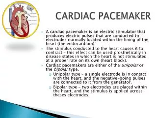
Permanent PACEMAKER Ventricular / Atrial channel transmits the Pacing impulse to the respective lead. Power source, Circuitry, Channels. Electrodes: Invasive / non invasive Uni /Bi polar
Designs of Electrodes • Invasive (epicardial or endocardial) or non-invasive (transcutaneous or transoesophageal) • Bipolar (>1 electrodes attached to myocardium) or unipolar (1electrode to heart and other to remote location) Ring Electrode Tip Electrode
Indication of Permanent Pacing • Sinus Node Dysfunction - Symptomatic diseases of impulse formation. • Sinus bradycardia, sinus pause or arrest, or sinoatrial block, chronotropic incompetence. • Often associated with paroxysmal SVTs. • 5O% implantation. • Atrial Fibrillation -Dual site atrial pacing to decrease intra-atrial conduction time.
Indications cont.. C. AV Block: Due to ischaemic / congenital / degenerative / inflammatory. • Block within HIS Purkinje system more malignant. • Class I - 3⁰ block with symptomatic bradycardia, documented asystole, escape rate <40 bpm, postop block unlikely to resolve, NM disease with block. • Class II — Asymptomatic 3⁰ block, asymptomatic Type II 2⁰ block, asymptomatic Type I 2 ⁰ block at or below bundle of HIS. D. Chronic BBB likely to progress to CHB.
Indications cont.. E. Miscellaneous • Hypertrophic Obstructive Cardiomyopathy, • Dilated cardiomyopathy, • Hypersensitive Carotid Sinus Syndrome & Neurogenic Syncope, • Cardiac Transplant, • Prolonged QT interval, • LV systolic dysfunction – biventricular / septal.
Generic Pacemaker Codes The North American Society of Pacing and Electrophysiology (NASPE) and British Pacing and Electrophysiology Group (BPEG) Pacemaker codes. NBG Pacemaker Codes:
Position III: Response(s) to Sensing • I (Inhibited): The chamber is paced unless intrinsic electrical activity is detected during the pacing interval. • T (Triggered): The pacing device will emit a pulse only in response to sensed event. Used for testing device. • D (Dual): Provides AV synchrony. Pacing device emit’s atrial pulse if no sensed atrial event takes place within timeframe, once an atrial event has occurred (native or paced), the pacing device will ensure that a ventricular event follows.
Position IV: Programmability very sensitive
Position V: Multisite Pacing • With 2002 revision of the NBG. the fifth column describes multisite pacing functionality . • Atrial multisite pacing might prevent atrial fibrillation. • Ventricular multisite pacing is an acceptable means of pacing patients with dilated cardiomyopathy .
Types of Pacing Modes • Asynchronous • AOO / VOO / DOO Asynchronous atrial / ventricular / atrioventricular sequential, fixed rate pacing, regardless to underlying cardiac rhythm. • Used safely in cases with NO ventricular activity. • Disadvantages: • Competes with patient’s intrinsic rhythm & results in induction of tachyarrythmias. • Continuous pacing wastes energy & decreases battery half-life.
Types of Pacing Modes cont.. • Single Chamber Atrial Pacing (AAI, AAT) • Atrial-only antibradycardia pacing. • Used sinus arrest and sinus bradycardia provided AV conduction is adequate. • Inappropriate for chronic AF & long ventricular pauses. • A single pacing lead with electrode in RA, senses intrinsic P wave & causes inhibition or triggering of pacemaker.
Types of Pacing Modes cont.. • Single Chamber Ventricular Pacing (VVI, VVT) • Ventricular-only antibradycardia pacing. • Indicated complete heart block with chronic atrial flutter, AF & long ventricular pauses. • Senses intrinsic R wave & thus inhibits or triggers pacemaker function. No atrial sensing; thus, no AV synchrony.
Types of Pacing Modes cont.. • Dual Chamber AV Sequential Pacing (DDD, DVI, DDI, VDD) • Two leads: unipolar or bipolar are used. Atrium is stimulated first , then after an adjustable PR interval ventricle is stimulated . • Preserve the normal atrioventricular contraction sequence. • Indicated AV block, carotid sinus syncope & sinus node disease.
DDD: After any sensed or paced atrial event, an intrinsic ventricular event must occur before expiration of AV timer. • Advantage: Generate sinus rhythm & beneficial where atrial contraction is important for ventricular filling (aortic stenosis). • Disadvantage: Pacemaker-mediated tachycardia (PMT) due to ventriculoatrial conduction, which triggers a ventricular depolarization leading to PMT. can be overcome by careful programming. • DDI: Ventricle is paced only when no intrinsic ventricular activity . Used inparoxysmal atrial fibrillation. • VDD: Used in patient with AV nodal dysfunction but intact SA node function.
a) DDD pacing in patient with normal SA node function and normal AV node function. b) DDD pacing in patient with normal SA node function and abnormal AV node function. c) DDD pacing in patient with abnormal SA node function and abnormal AV node function.
British pacing and electro physiology group recommended pacemaker modes.
PACEMAKER FAILURE • Pacemaker failure has three etiologies: 1) Failure to capture: • Myocardial ischemia/infarction, • Acid-base disturbance, • Electrolyte abnormalities, • Abnormal antiarrhythmic drug levels. • External pacing might further inhibit pacemaker. 2) Lead failure, 3) Generator failure.
ICD Implantable Cardioverter Defibrillator • Battery powered device to deliver sufficient energy to terminate VT / VF. • Superior to anti-arrhythmic therapy in preventing death in malignant ventricular tachy-dysrhythmias.
ICD use rate criteria to detect VF • Have VT detection algorithms based on RR interval • Tachycardia with cycle length less than VF detection interval initiates VT therapy
Indications for ICDs • Ventricular Tachycardia • Ventricular Fibrillation • Brugada Syndrome (RBBB, ST-segment elevation in V1 to V3) • Arrhythmogenic RV Dysplasia • Long Q-T Syndrome • Hypertrophic cardiomyopathy • Prophylactic use in patient who has cardiomyopathy with EF ≤ 35% (SCD-HeFT) & Post-MI patients with EF ≤ 30% .
Effect of Magnet • Each PM/ICD is programmed to respond in a specific manner to magnet placement. • Magnet usually result in pacemaker to switch to asynchronous mode. • A slower than expected (≥10%) magnet rate is a marker for battery depletion. • Magnet never turn off pacemaker. • ICD will be inhibited to deliver antitachycardia therapy when magnet is applied. • Pacemaker function of ICD is not inhibited.
Magnet application to a VVI pacemaker. Magnet application Normal sinus rhythm with normal AV conduction Fixed rate , ventricular pacing
Anesthetic Considerations Practice Advisory for the Perioperative Management of Patients with Cardiac Rhythm Management Devices, ASA , Anesthesiology 2005; 103:186–198. Preoperative Evaluation • Establish that patient has a cardiac rhythm management device (CRMD). • Detailed focused history (patient interview, medical records review, review CXR, ECG, SE). • Physical examination (check for scars, palpate for device). • Define the type of CRMD. • Obtain manufacturer’s identification card. • Get CXR if no other data available.
Determine dependency on pacing function. • H/o symptomatic bradyarrhythmia resulting in CRMD implantation. • H/o successful atrioventricular nodal ablation. • Inadequate escape rhythm at lowest programmable pacing rate. • Determine CRMD function. • Interrogate device (consult cardiologist or PM-ICD service). • Determine whether the device will capture when it paces. • Consider contacting the manufacturer for perioperative recommendations.
Preoperative Preparation • Determine whether EMI is likely to occur during the procedure. • Determine whether reprogramming pacing function to asynchronous mode or disabling rate responsive function is advantageous. • Suspend antitachyarrhythmia functions if present. • Advise surgeon to consider use of a bipolar electrocauteryor ultrasonic (harmonic) scalpel. • Temporary pacing and defibrillation equipment should be immediately available. • Evaluate the possible effects of anesthetic techniques and of procedure on CRMD function.
Intraoperative Management • Monitor operation of CRMD. • ECG per ASA standard. • Monitor peripheral pulse (manual pulse palpation, pulse oximeterplethysmogram, arterial line). • HOCM patients need beat-to-beat cardiac output monitoring. • Though anaesthetic techniques dont influence CRMD function, the anaesthetic-induced changes (HR / rhythm / ischaemia) may induce unexpected CRMD responses
Factors affecting pacing thresholds Increase • 1-4 weeks after implantation • Myocardial ischaemia/infaction • Hypothermia, hypothyroidism • Hyperkalaemia, acidosis/alkalosis • Antiarrythmics • Severe hypoxia & hypoglycaemia. Decrease • Increased catecholamines • Stress, anxiety • Sympathomimetic drugs • Anticholinergics • Glucocorticoides • Hyperthyroidism • Hypermetabolic status.
Manage potential CRMD dysfunction due to EMI. • Electrocautery. • Assure that electrosurgical receiving plate is positioned so that current pathway does not pass through CRMD. • Advise surgeons to avoid proximity of cautery to PM or leads. • Advise surgeons to use short, intermittent, and irregular bursts at the lowest feasible energy levels. • Advise surgeons to reconsider use of bipolar electrocautery system. • Radiofrequency ablation. • Advise surgeons to avoid direct contact between the ablation catheter and PM and leads. • Advise surgeons to keep radiofrequency current path as far away from PM and leads.
Lithotripsy. • Advise surgeons to avoid focusing the lithotripsy beam near pulse generator. • If the lithotripsy system triggers on the R wave, consider preoperative disabling of atrial pacing. • MRI. • MRI is generally contraindicated in patients with CRMDs. • If MRI must be performed, consult with the ordering physician, cardiologist, radiologist and CRMD manufacturer.
Radiation therapy. • Radiation therapy can be safely performed. • Surgically relocate the CRMD if the device will be in the field of radiation. • Electroconvulsive therapy. • No significant damage if CRMD disabled • Consult with the ordering physician, cardiologist, CRMD manufacturer.
Emergency defibrillation or cardioversion. • For a patient with ICD and magnet-disabled or programming-disabled therapies: • Advise surgeons to terminate all sources of EMI while magnet is removed. • Remove the magnet or reprogramming to reenableantitachycardia therapies. • Observe patient & the monitors for appropriate CRMD therapy. • If the above activities do not restore ICD function, proceed with emergency external defibrillation or cardioversion.
For external defibrillation: • If its technically impossible, defibrillate patient in the quickest possible way and be prepared to provide pacing through other routes. Lt pectoral implant showing acceptable anterior-apex pad placement Left or right or left pectoral implant showing recommended anterior-posterior pad placement View of anterior-posterior pad placement
Postoperative Management • Continuously monitor HR & rhythm. • Have backup pacing & defibrillation equipment available throughout the immediate postoperative period. • Interrogate and restore CRMD function in the immediate postoperative period. • Restore all antitachyarrhythmic therapies in ICDs. • Assure that all other settings of the CRMD are appropriate.
References • Practice Advisory for the Perioperative Management of Patients with Cardiac Rhythm Management Devices: Pacemakers and Implantable Cardioverter Defibrillators, ASA , Anesthesiology 2005; 103: 186–198. • T. V. Salukhe, D. Dob and R. Sutton, Pacemakers and defibrillators: anaesthetic implications, Br J Anaesth 2004; 93: 95-104. • ACC/AHA/HRS 2008 Guidelines for Device-Based Therapy of Cardiac Rhythm Abnormalities, J. Am. Coll. Cardiol. 2008; 51; e1-e62 (http://content.onlinejacc.org/cgi/content/full/51/21/e1) • Kaplan’s Cardiac Anesthesia; 5th edition. • Miller’s Anesthesia; 7th edition. • Stoelting’s Anesthesia & Co-existing Disease; 5th edition.