Download
1 / 5
50 likes | 179 Vues
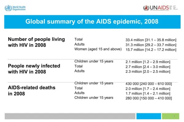
This case study details the prenatal diagnosis and management of a fetus with congenital heart defects. A comprehensive ultrasound was conducted at 25 weeks of gestation, followed by a heart CT scan before the delivery of a baby boy weighing 2780g. The infant was delivered vaginally at 38 weeks and required admission to the neonatal intensive care unit for echocardiography and cardiac surgery. The findings included abnormal vessels and collateral circulation assessed via Doppler and CT imaging. Key questions address sonographic abnormalities, diagnosis, and details of collateral vessels.

E N D
HKSUM CME program 2009 December Acknowledgement: Prof. Hye-Sung Won M.DDepartment of Obstetrics and Gynecology University of Ulsan College of Medicine, Asan Medical Center, Seoul, Korea Sponsored by Medison company
Case A prenatal detailed ultrasound examination was performed at 25 weeks’ gestation. A heart CT was subsequently performed. At 38 weeks’ gestation, a baby boy of 2780g with AS 7 at 1 minute and 8 at 5 minutes was delivered vaginally. The baby was admitted to neonatal intensive care unit. Echocardiography, and cardiac surgery was subsequently performed.
LVOT FCV B-mode Doppler Abnormal vessels 4CV VSD Lt Lt RV LV Ao 77.3° Desc.Ao
LV RV LA RA Color Doppler PA Ao SVC Heart computed tomography. a. Three collateral vessels (C1-C3) from descending thoracic aorta. b. schematic picture of collateral vessels.
Questions • 1. What are the sonographic abnormalities? • 2. What is the likely diagnosis? • 3. What are the three collateral vessels as shown on CT?