Download

1 / 29
480 likes | 1.31k Vues
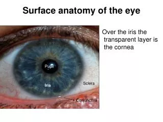
Gross Anatomy of the Eye. Cornea at anterior Light passes to lens Retina at posterior sensory tissue sensory cells: rods and cones. Sup. Lat. Med. Inf. 1. Cornea 2. Lens 3. Iris 4. Sclera 5. Macula 6. Optic Nerve Head 7. Retinal vessels 8. Vortex Veins. Looking at the Retina.

E N D
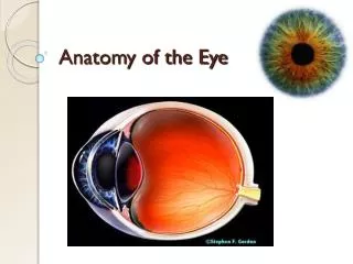
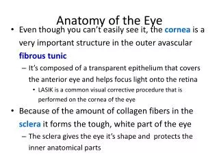
Gross Anatomy of the Eye • Cornea at anterior • Light passes to lens • Retina at posterior • sensory tissue • sensory cells: rods and cones
Sup. Lat. Med. Inf. 1. Cornea 2. Lens 3. Iris 4. Sclera 5. Macula 6. Optic Nerve Head 7. Retinal vessels 8. Vortex Veins
Looking at the Retina Macula- 3 by 5 mm area at the posterior pole of the eye Fovea- in center of macula, free of blood vessels contains only cone cells
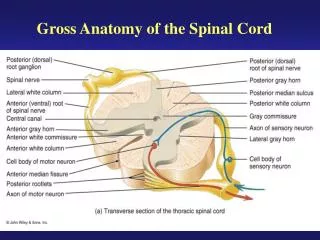
Back of the Eye Front of the Eye
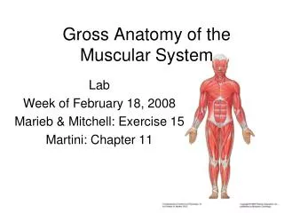
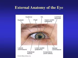
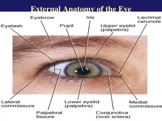
Extrinsic Eye Musculature For moving eye within its socket. 6 muscles per eyeball Innervated by 3 Cranial nerves
YAW Eye Movements Yaw: gaze shifts L/R Pitch: gaze shifts up/down Roll: eye rotates around line of gaze (torsion) • Adduction: shifting gaze toward midline • Abduction: shifting gaze laterally PITCH ROLL
Extraocular Muscles Anterior View of Left Orbit
Sup. Post. Ant. Inf. 3 branch of CN III to Inf Obl. 5 Sup. Rectus 7 Inf Rectus 8 Optic Nerve 10 Abducens Nerve 11 Oculomotor Nerve (CN III)
Ocular Musculature Superior Rectus (SR) Inferior Rectus (IR) Lateral Rectus (LR) Medial Rectus (MR) Superior Oblique (SO) Inferior Oblique (IO)
Cranial Nerves III, IV, and VI • III - Oculomotor • IV - Trochlear • VI - Abducens
III (Oculomotor) innervates: 1) Medial rectus 2) Superior rectus 3) Inferior rectus 4) Inferior oblique Levator palpebrae sup Pupillary sphincter Ciliary muscle
IV (Trochlear) innervates: • Superior oblique
VI (Abducens) innervates • Lateral rectus.
Proprioceptive info from eye muscles • comes through Trigeminal nerve.
Eye Movements • Saccades—rapid shift in gaze • Pursuit—stabilize image of moving object • Fixation—stabilize image of still object • VOR—stabilize image during head motion • OKN—backup for when VOR decays to cont’d head rotation • Vergent movements—change depth of focus • Accommodation-- automatic changes to see at different distances which is chiefly brought about by changes in the convexity of the lens. Horizontal vergence and accommodation normally occur together. The two responses are accompanied by an appropriate change in pupil diameter. The three concomitant changes are known as the near-triad response.
Cortical Areas: Oculomotor Control • Occipital Eye Fields (areas 18 and 19) • Frontal Eye Fields (area 8) • Temporal Eye Fields (area 22)
VOR Pathways • Vestibular nuclei • Abducens N. • Median Longitudinal Fasciculus • Trochlear N. • Oculomotor N.
Saccades Pause cells inhibit Burst Neurons which stimulate: III & VI (horizontal) or III & IV (vertical)