Download
1 / 44
470 likes | 1.3k Vues
Chronic Obstructive Airways Disease. Gerard Flaherty B.Sc.(Hons.), M.B., B.Ch., B.A.O., M.R.C.P.I. Specialist Registrar in General Medicine/Endocrinology. Lecture Outline. Definition of COAD Diagnostic criteria Classification of severity Epidemiology Chronic bronchitis Emphysema

E N D
Chronic Obstructive Airways Disease Gerard Flaherty B.Sc.(Hons.), M.B., B.Ch., B.A.O., M.R.C.P.I. Specialist Registrar in General Medicine/Endocrinology
Lecture Outline • Definition of COAD • Diagnostic criteria • Classification of severity • Epidemiology • Chronic bronchitis • Emphysema • Bronchiectasis • Bronchial asthma
Definition of COAD • COAD is a disease state characterised by airflow limitation that is not fully reversible. The airflow limitation is usually both progressive and associated with an abnormal inflammatory response of the lungs to noxious particles or gases.
Epidemiology of COAD • Prevalence and morbidity data greatly underestimate the total burden of COAD. • Prevalence of 9.34/1,000 in men and 7.33/1,000 in women (Global Burden of Disease Study, 1990). • Morbidity increases with age and is greater in men than in women. • Currently the 4th leading cause of death in the world. • Economic burden: per capita cost of COAD in UK of US$65 (1996).
Host factors Alpha-1-antitrypsin deficiency Asthma and airway hyperresponsiveness Disordered lung development Environmental factors Tobacco smoke Occupational dusts/chemicals Air pollution Childhood infections Lower socioeconomic status Risk Factors for COAD
Diagnosis of COAD • Considered in patients with cough, sputum production, or dyspnoea +/- risk factors. • Confirmed by spirometry. • FEV1/FVC <70% + postbronchodilator FEV1 <80% of predicted value. • A low peak expiratory flow has poor specificity for the diagnosis of COAD.
Spirometry • Normal flow-volume loop • Flow-volume loop in severe COAD
Respiratory Failure • Results when gas exchange is inadequate, resulting in hypoxia with PaO2 <8kPa. • Type I: Hypoxia with normal/low PaCO2. • Caused by ventilation/perfusion mismatch, e.g., Life-threatening asthma, Emphysema. • Type II: Hypoxia with hypercapnia (PaCO2 >6.5kPa). • Caused by alveolar hypoventilation, e.g., Chronic bronchitis. • Oxygen therapy must be controlled in type II respiratory failure due to the risk of further hypercapnia if the hypoxic drive to ventilation is abrogated by excessive oxygen.
Chronic Bronchitis • Persistent cough with sputum production for at least 3 months in ≥ consecutive years • Simple / Chronic asthmatic / Obstructive • Most frequent in middle-aged men • Higher incidence in urban dwellers • May coexist with emphysema • Presents with exertional dyspnoea and frequent respiratory tract infections
Chronic BronchitisPathogenesis • Chronic irritation by inhaled substances • Respiratory infections • 4-10 times more common in heavy smokers • Hypersecretion of mucus in large airways • Submucosal gland hypertrophy • Increase in goblet cells in small airways • Excessive mucus → airway obstruction • Cigarette smoke predisposes to infection
Gross Hyperaemia and oedema of mucous membranes Excessive secretions +/- casts Histology Hypertrophy of submucosal smooth muscle Hyperplasia of submucosal glands of trachea/bronchi Increased Reid index (>0.4) Squamous metaplasia and dysplasia of epithelium Loss of ciliary activity Narrowing of bronchioles +/- bronchiolitis obliterans Chronic BronchitisMorphology
Chronic BronchitisComplications • ↓PaO2 ↑PaCO2 ….Respiratory failure • Respiratory tract infections (H. influenzae, Strep. Pneumoniae) • Pulmonary hypertension → Cor pulmonale • Atypical metaplasia / dysplasia of respiratory epithelium….Bronchogenic carcinoma
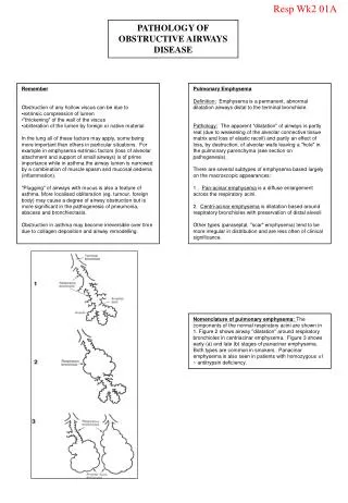
Emphysema • Abnormal permanent enlargement of the airspaces distal to the terminal bronchiole, accompanied by destruction of their walls • Types: (1) centrilobular (2) panlobular (3) paraseptal (4) irregular • Associated with heavy cigarette smoking
Emphysema • Presents with progressive dyspnoea, reduced exercise tolerance, wheeze and weight loss • Signs of hyperinflation • Loss of elastic support → Airways collapse on expiration → Obstructive pattern on spirometry • Respiratory acidosis • Cor pulmonale (rarely) • Pneumothorax
EmphysemaPathogenesis • Protease-antiprotease hypothesis • Homozygous alpha-1-antitrypsin deficiency is associated with panlobular emphysema, especially in smokers • Emphysema results from the destructive effect of high protease (elastase) activity in patients with low antiprotease activity • Smoking stimulates release of elastase from neutrophils
Gross Panlobular: resp. bronchiole → terminal alveoli. Lower > Upper lobes Centrilobular → respiratory bronchioles. Upper > Lower Paraseptal → distal acinus. Upper > Lower Irregular → associated with scarring, e.g., Tuberculosis Histology Destruction of septal walls Fusion of adjacent alveoli Blebs / bullae Compressed blood vessels +/- Bronchitis EmphysemaMorphology
Bronchiectasis • A chronic necrotising infection of the bronchi and bronchioles associated with abnormal permanent dilation of these airways • Presents with cough, productive of large amounts of foul-smelling, purulent sputum, hamoptysis and digital clubbing • Pooled secretions in lower lobes → respiratory tract infections
BronchiectasisAetiology • Bronchial obstruction Tumour, foreign bodies, mucous impaction Asthma / Chronic bronchitis • Infection Tuberculosis, Staphylococcus aureus, ABPA, Measles, Pertussis • Congenital Congenital bronchiectasis, Cystic fibrosis, Intralobar sequestration, Immunodeficiency, Kartagener’s syndrome, Yellow nail syndrome
BronchiectasisPathogenesis • Bronchial obstruction → Atelectasis • Dilatation of walls of patent airways • Infection → Bronchial wall inflammation → weakened walls → further dilation • Cystic fibrosis: squamous metaplasia with impaired mucociliary action, infection, necrosis of bronchial and bronchiolar walls • Kartagener’s syndrome: absent dynein arms in cilia → lack of ciliary activity
Gross Usually both lower lobes May be localised Dilated airways Cylindroid Fusiform Saccular Cystic pattern on cut surface of lungs Histology Acute and chronic inflammation Desquamation of epithelium Necrotising ulceration Squamous metaplasia Necrosis → lung abscess Fibrosis BronchiectasisMorphology
BronchiectasisComplications • Pneumonia • Lung abscess • Empyema • Septicaemia • Cor pulmonale • Metastatic cerebral abscesses • Secondary Amyloidosis
Bronchial Asthma • Hyper-reactive bronchioles, leading to episodic, reversible bronchoconstriction • Bronchospasm → cough, dyspnoea, wheeze • Status asthmaticus may be fatal • Types: (1) Extrinsic (eg, atopic, occupational, ABPA*) – type 1 reaction (2) Intrinsic (eg, respiratory infections, aspirin, exercise)
Atopic Asthma Exposure of presensitised IgE-coated mast cells to antigen causes acute immediate response Chemical mediators (histamine, cytokines, PAF, leukotrienes) Late-phase reaction Nonatopic Asthma Virus-induced inflammation of respiratory mucosa lowers threshold of vagal receptors to irritants Aspirin-sensitive asthma a/w rhinitis/nasal polyps Occupational asthma Bronchial AsthmaPathogenesis
Bronchial AsthmaMorphology Gross • Overdistended lungs • Mucous plugs occlude bronchioles Histology • Thickened basement membrane • Oedema • Inflammatory infiltrate • Numerous eosinophils • Hyperplasia of submucosal glands • Bronchial wall muscle hypertrophy • Curschmann’s spirals • Charcot-Leyden crystals (eosinophil membrane protein)
Bronchial AsthmaComplications • Status asthmaticus • Pneumothorax • Bronchiectasis • Cor pulmonale