Download
1 / 50
570 likes | 783 Vues
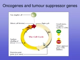
Cancer pathogenesis Oncogenes Tumor suppressor genes Invasion and metastasis Epithelial-mesenchymal transition (EMT) Cancer stem cells. Jimin Shao shaojimin@zju.edu.cn. 1. Oncogene.

E N D
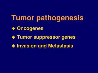
Cancer pathogenesis • Oncogenes • Tumor suppressor genes • Invasion and metastasis • Epithelial-mesenchymal transition (EMT) • Cancer stem cells Jimin Shao shaojimin@zju.edu.cn
1. Oncogene • Oncogene: a gene that when mutated or expressed at abnormally-high levels contributes to converting a normal cell into a cancer cell. • The identification of oncogene abnormalities has provided tools for the molecular diagnosis and monitoring of cancer. • Oncogenes represent potential targets for new types of cancer therapies. • Cellular oncogene (c-onc): -- proto-oncogene:in normal physiologic version -- oncogene:altered in cancer • Viral oncogene (v-onc)
Fuctions of proto-oncogenes Proto-oncogenes at all levels of the various signal transduction cascades that control cell growth, proliferation, and differentiation: • extracellular proteins:growth factors, membrane proteins: cell surface receptors, • cellular proteins: relay signals • proteins innucleus: Transcription Factors, promoters of cell cycle
Mechanisms of Oncogene Activation 1. Gene amplification, e.g.myc, CCND1 2. Point mutation, e.g.ras, 3. Chromosomal rearrangement or translocation -- transactivation of proto-onc -- fusion genes, e.g.abl-bcr 4. Viral insertion activation, e.g.c-Myc
Ras • Locates on chromosome 11, codes for a protein with GTPase activity • relays signals by acting as a switch:When receptors on the cell surface are stimulated, Ras is switched on and transduces signals that tell the cell to grow. If the cell-surface receptor is not stimulated, Ras is not activated and so the pathway that results in cell growth is not initiated. • mutated in about 30% of cancers so that it is permanently switched on, telling the cell to grow regardless of whether receptors on the cell surface are activated or not.
Ras relays signals from the cell surface receptors to the nucleus • Ras relays signals by acting as a switch
2. Tumor suppressor genes TSGs: genes that sustain loss-of function mutations in the development of cancer
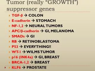
TSGs: functions and tumor associations The biology of cancer (2nd edition), RA Weinberg, 2013
Mechanism for the inactivation of TSGs • Mutation: point mutation or frameshift mutation, p53 • Deletion: LOH or homozygous deletion, Rb • Viral oncoprotein inactivation: p53, Rb • Promoter hypermethylation, histone modification changes: p16
RB:Cell Cycle Controller Rb regulates G1/S transition
P53: Function as gatekeeper Inactivation of p53 in cancer • LOH on 17p13 in a number of tumors • Point mutation on exon 5-8 “hot-spot” (Dominant negative mutation) • MDM2 negative regulation • viral-oncogene products inactivation
(1) Introduction • Invasion and Metastasis: a hallmark of malignancy, occurs in four steps: • Growth at primary site and angiogenesis • Tumor cell invasion • Lymphatic and hematogenous metastasis • Growth at secondary site and angiogenesis
Up to 70% of patients with invasive cancer have overt or occult metastases at diagnosis. Acquisition of the invasive and metastatic phenotype is an early event in cancer progression. Millions of tumor cells are shed daily into the circulation. Less than 0.01% of circulating tumor cells successfully initiate a metastatic focus. Circulating tumor cells can be detected in patients who do not develop overt metastatic disease. Angiogenesis is a ubiquitous and early event that is necessary for and promotes metastatic dissemination.
(2) Mechanisms involved in cancer cell invasion 1) Loss of cell-to cell cohesive forces: Decreased cellular adhesion 2) Secretion of ECM-degrading enzymes: Degradation of ECM 3) Active Locomotion: Abnormal or increased cellular motility 4) Protein kinases 5) Tumor angiogenesis 6) Metastasis-related genes
1) Loss of cell-to cell cohesive forces: Cell adhesion molecules (CAMs): the membrane proteins that mediate selective adhesion between cells, and between cells and extracellular matrix (ECM). • E-cadherin: Expression↓ Loss of cell-cell adhesion,Increased cell motility • Integrins:Expression↓→↑ heterodimer (alfa-beta subunits) the ligands: ECM components, collagens (I,IV), laminin (LN), fibronectin (FN), etc. • Immunoglobin superfamily: VCAM-1, ICAM-1, CEA, DCC, etc • Selectins • CD44 variants
2) Secretion of ECM-degrading enzymes • Matrix Metalloproteinases (MMPs):~20 • Tissue inhibitors of metalloproteinases (TIMPs): ~4 • Plasminogen Activators (PAs) : urokinase-type (uPA), tissue-type PA (tPA) • PA inhibitors (PAIs): ~3 Metastasis-associated proteinases Cell invasion of the ECM
uPA¯uPAR-initiated signal transduction and consequences Plasminogen/Plasmin System
3) Active Locomotion • Autocrine motility factor (AMF), Autotaxin (ATX), • Cytoskeletal proteins • E- cadherin • Growth factors and receptors, • ECM components (laminin, LN, etc) 4) Protein Kinases • GPCR/RTK/CK-R/CAM-R---Rho GTPases ---signalling---cell mobility • Integrins---FAK (focal adhesion kinase) ---signalling---cell mobility
5) Tumor angiogenesis factors (TAFs)Models of Tumour Angiogenesis
Blocking Blood Supply to Tumors Many solid tumors are dependent on the growth of new blood and lymphatic vessels to grow and survive. In the past 10 years, the FDA has approved 10 antiangiogenic agents. The newest member of this growing class of therapeutics is ramucirumab (Cyramza). It was approved by the FDA for the treatment of metastatic gastric cancer and gastroesophageal junction adenocarcinoma in April 2014. Ramucirumab is also being tested in numerous clinical trials as a potential treatment for other types of cancer, such as NSCLC.
Metastasis-suppressor genes: 6) Metastasis-enhancing genes:Oncogenes,CD44, Integrinβ1, CEA, MMP2, u-PA, etc
(3) Metastasis Therapeutic Targets and Agents A. Targeted Therapeutics Target Example Agents Effects Growth factors C225 (anti-EGFR) Block growth factor signaling Tyrphostins (anti-RTK) Cell adhesion Anti-avb3 (Vitaxin) Blocks endothelial cell avb3 peptidomimetics interaction with matrix may regulate MP activation Proteolysis MMPIs uPAR-I Blocks degradation of matrix, blocks activation of proteases, growth factors Motility Taxanes Blockade of microtubule cycling
B. Signal Inhibitors: Blockade of signals necessary for angiogenesis , invasion, and metastasis Agent Target Activity CAI Calcium influx Inhibits adhesion, motility, angiogenesis Squalamine Inhibits NHE-3 Anti-angiogenic PI3K inhibitors Inhibit motility, proliferation, promote MAPK inhibitors Inhibit invasion, proliferation
[J Clin Invest. 2009;119(6):1420-8. doi: 10.1172/JCI39104] 4. Epithelial-mesenchymal transition (EMT) (1) EMT有三种形式: • I型EMT(上皮细胞-间质细胞转换):胚胎发育; • 发育异常,畸形(先天性瓣膜性心脏病) • II型EMT(上皮细胞-成纤维细胞转换):创伤修复,瘢痕形成; • 不愈合,过度愈合(纤维化性疾病) • III型EMT (肿瘤上皮-间质细胞转换) 肿瘤侵袭转移;耐药,酸中毒抵抗,凋亡抵抗;干细胞样特征
Epithelial-mesenchymal plasticity allows cancer cells to undergo functional adaptations during the invasion-metastasis cascade W. L. Tam, R. A. Weinberg, Nat Med 19, 1438 (Nov, 2013)
(3) EMT Signaling Pathways J. P. Thiery, H. Acloque, R. Y. Huang, M. A. Nieto, Cell 139, 871 (2009)
5. Cancer stem cells • tumorigenesis • metastasis • drug resistance • relapse Z. Yu, Y. Li, H. Fan, Z. Liu, R. G. Pestell, Front Genet 3, 191 (2012)
The hypothesis of miPS differentiation when exposed to normal or malignant niche. miPS cells should be induced to some kinds of progenitor cells, such as hematopoietic cells and neural stem cells, differentiating into various phenotypes, such as macrophage, monocytes, neural cells, cardiac cells and pancreatic b- cells, when exposed to the normal niche. We hypothesized that CSCs may also be derived from miPS cells only when exposure to a malignant niche. [Chen L, Kasai T, Li Y, et al. A model of cancer stem cells derived from mouse induced pluripotent stem cells. PLoS One. 2012;7(4):e33544. doi:10.1371/journal.pone.0033544.g001]
Cancer Cell Metastasis Cascade Chaffer CL, Weinberg RA. A Perspective on Cancer Cell Metastasis. Science 331, 1559 (2011)
思考题: 1. 简述原癌基因和抑癌基因概念,以及原癌基因激活、抑癌基因失活的机制。 2. 简述肿瘤转移的基本过程和机制。 3. 简述肿瘤干细胞、肿瘤微环境、EMT概念,