Download
1 / 77
1.14k likes | 3.01k Vues
Tumor pathogenesis Oncogenes Tumor suppressor genes Invasion and Metastasis. Introduction. Carcinogensis is multistep process involving the multiple genetic changes including the activation of cooperating oncogenes and the inactivation of tumor suppressors in somatic cells. 2.

E N D
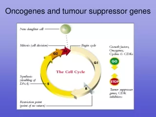
Tumor pathogenesis • Oncogenes • Tumor suppressor genes • Invasion and Metastasis
Introduction • Carcinogensis is multistep process involving the multiple genetic changes including the activation of cooperating oncogenes and the inactivation of tumor suppressors in somatic cells 2
Usually, a single oncogene is not enough to turn a normal cell into a cancer cell, and many mutations in a number of different genes may be required to make a cell cancerous.
Figure 2. Intracellular Signaling Networks Regulate the Operations of the Cancer Cell. An elaborate integrated circuit operates within normal cells and is reprogrammed to regulate hallmark capabilities within cancer cells. Separate subcircuits, depicted here in differently colored fields, are specialized to orchestrate the various capabilities. At one level, this depiction is simplistic, as there is considerable crosstalk between such subcircuits. In addition, because each cancer cell is exposed to a complex mixture of signals from its microenvironment, each of these subcircuits is connected with signals originating from other cells in the tumor microenvironment, as outlined in Figure 5. (Hanahan D, Weinberg RA. Hallmarks of Cancer: The Next Generation. Cell 2011, 144:646)
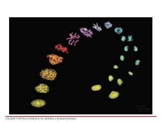
Michael R. Stratton. Exploring the Genomes of Cancer Cells: Progress and Promise. Science 331, 1553 (2011).
Michael R. Stratton. Exploring the Genomes of Cancer Cells: Progress and Promise. Science 331, 1553 (2011).
Oncogene Concept: An oncogene is a gene that when mutated or expressed at abnormally-high levels contributes to converting a normal cell into a cancer cell. • Cellular oncogene (c-onc): --- proto-oncogene (proto-onc):in normal physiologic version --- Oncogene:altered in cancer • Viral oncogene (v-onc)
Fuctions of proto-oncogenes • Proto-oncogenes have been identified at all levels of the various signal transduction cascades that control cell growth, proliferation and differentiation: • extracellular proteins function as growth factors, • membrane proteins as cell surface receptors • cellular proteins that relay signals • proteins innucleus, which activate the transcription and promote the cell cycle • This signaling process involves a series of steps that: • begin from the extracellular environment to cell membrane; • involve a host of intermediaries in the cytoplasm; • end in the nucleus with the activation of transcription factors that help to move the cell through its growth cycle.
Classification of proto-oncogenes • Growth factors, e.g. V-sis, PDGF-b, int-2 • Receptor Tyrosine Kinases,e.g.Her-2/neu/ erbb2, • Membrane Associated Non-Receptor Tyrosine Kinases, e.g.src, Lck • G-Protein Coupled Receptorse.g. Mas • Membrane Associated G-Proteins,e.g. Ras • Serine/Threonine Kinases e.g. Raf • Nuclear DNA-Binding/Transcription Factors, e.g. myc, fos • Others • Apoptosis regulators, e.g. Bcl-2, • Regulators of cell cycle, e.g.Cyclin D1, CDK4
Mechanisms of Oncogene Activation • Gene amplification, e.g.myc, CCND1 • Point mutation, e.g.ras, • Chromosomal rearrangement or translocation • the transcriptional activation of proto-onc. • the creation of fusion genes, e.g.abl-bcr • Viral insertion activation, e.g.c-Myc
Amplification Translocation
CHROMOSOMAL REARRANGEMENTS OR TRANSLOCATIONS Neoplasm Translocation Proto-oncogene Burkitt lymphoma t(8;14) 80% of cases c-myc1 t(8;22) 15% of cases t(2;8) 5% of cases Chronic myelogenous t(9;22) 90-95% of cases bcr-abl2 leukemia Acute lymphocytic t(9;22) 10-15% of cases bcr-abl2 Leukemia 1c-myc is translocated to the IgG locus, which results in its activated expression 2bcr-abl fusion protein is produced, which results in a constitutively active abl kinase
GENE AMPLIFICATION Oncogene Amplification Source of tumor c-myc ~20-fold leukemia and lung carcinoma N-myc 5-1,000-fold neuroblastoma retinoblastoma L-myc 10-20-fold small-cell lung cancer c-abl ~5-fold chronic myoloid leukemia c-myb 5-10-fold acute myeloid leukemia colon carcinoma c-erbB ~30-fold epidermoid carcinoma K-ras 4-20-fold colon carcinoma 30-60-fold adrenocortical carcinoma
Ras • Locates on chromosome 11, codes for a protein with GTPase activity • relays signals by acting as a switch:When receptors on the cell surface are stimulated, Ras is switched on and transduces signals that tell the cell to grow. If the cell-surface receptor is not stimulated, Ras is not activated and so the pathway that results in cell growth is not initiated. • mutated in about 30% of human cancers so that it is permanently switched on, telling the cell to grow regardless of whether receptors on the cell surface are activated or not.
Her2/neu/erbB-2 • This gene was discovered by three different groups. That is why it has three different names. • It is a member of EGFR superfamily, also be a receptor tyrosine kinases • Dr. Slamon (UCLA) described the role of Her2/neu in breast cancer and ovarian cancer. • Overexpression, amplification, rare translocations • No ligand is known
Ras relays signals from the cell surface receptors to the nucleus Ras relays signals by acting as a switch
Prospect • A breakthrough for our understanding of the molecular and genetic basis of cancer • Provided important knowledge concerning the regulation of normal cell proliferation, differentiation, and programed cell death. • The identification of oncogene abnormalities has provided tools for the molecular diagnosis and monitoring of cancer. • Oncogenes represent potential targets for new types of cancer therapies.
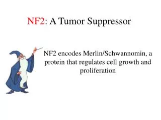
Tumor suppressor genes Concept: genes that sustain loss-of function mutations in the development of cancer
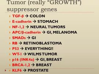
TSGs Transcriptional factor: p53, WT1, Direct transcription regulator: Rb, APC Inhibitor of cell cylcle kinase: p16INK4A, p19ARF, Cell structural components: NF2 Phosphatase: PTEN Potential mediator of mRNA processing: VHL Components involved in DNA repair: MSH2, MLH1, BRCA1, p53
TUMOR SUPPRESSOR GENES Disorders in which gene is affected Gene (locus) Function Familial Sporadic DCC (18q) cell surface unknown colorectal interactions cancer WT1 (11p) transcription Wilm’s tumor lung cancer Rb1 (13q) transcription retinoblastoma small-cell lung carcinoma p53 (17p) transcription Li-Fraumeni breast, colon, syndrome & lung cancer BRCA1(17q) transcriptional breast cancer breast/ovarian tumors BRCA2 (13q) regulator/DNA repair
Mechanism for the inactivation of TSGs • Mutation: point mutation or frameshift mutation, p53 • Deletion: LOH (loss of heterozygosity) or homozygous deletion, Rb • Viral oncoprotein inactivation: p53, Rb • Promoter hypermethylation, histone modification changes: p16
Rb regulates G1/S transition Rb inactivation by viral oncoprotein
RB RB RB LOH RB Mutation KNUDSON TWO HIT HYPOTHESIS IN SPORADIC CASES Normal Cells RB RB Inactivation of a tumor suppressor gene requires two somatic mutations. Tumor cells
Bax P53 Function as gatekeeper • Inactivation of p53 in cancer • LOH on 17p13 in a number of tumors • Point mutation on exon 5-8 “hot-spot” (Dominant negative mutation) • MDM2 negative regulation • viral-oncogene products inactivation
The process of metastasis consists of sequential linked steps • Growth at primary site and angiogenesis • Tumor cell invasion • Lymphatic and hematogenous metastasis • Growth at secondary site and angiogenesis
Mechanisms involved in tumor cell invasion • 1.Loss of cell-to cell cohesive forces • 2. Secretion of ECM-degrading enzymes • 3. Active Locomotion • 4. Tumor angiogenesis • 5. Metastasis-related genes
5. Metastasis-enhancing Genes:Oncogenes,CD44, Integrinβ1, CEA, MMP2, u-PA, etc
1. Loss of cell-to cell cohesive forces: • Cell adhesion molecules (CAMs): • 细胞粘附分子:介导细胞之间或细胞与ECM之间的选择性粘附。 • E-cadherin: Expression↓ • Loss of cell-cell adhesion,Increased cell motility • Integrins:Expression↓→↑ • Immunoglobin superfamily:NCAM, VCAM-1,CEA, DCC, etc • Selectins • CD44 variants
2. Secretion of ECM-degrading enzymes • Matrix Metalloproteinases (MMPs):~20 • Tissue inhibitors of metalloproteinases (TIMPs): ~4 • Plasminogen Activators (PAs) :urokinase-type, tissue-type PA • PA inhibitors (PAIs): ~3 Metastasis-associated proteinases
The MMP family (at least 23 members) Classification
基质金属蛋白酶 • 间质胶原酶(Interstitial Collagenase ),如MMP-1、MMP-5、MMP-8、 MMP-13等,作用底物主要为间质胶原 Ⅰ、Ⅱ、Ⅲ、Ⅶ、Ⅹ型胶原,但不能降解明胶和Ⅳ型胶原。 • 明胶酶(Gelatinase )又称Ⅳ型胶原酶,如MMP-2、 MMP- 9,作用底物主要是Ⅳ型胶原和明胶,还可以降解Ⅵ、Ⅶ、Ⅷ和Ⅹ型胶原,但不能降解间质胶原。 • 基质溶解素(Stromelysin),如MMP-3、 MMP-7、 MMP-10 和MMP-11等,作用底物主要是基质中的蛋白多糖和糖蛋白,如纤维连接蛋白(FN)、层黏连蛋白(LN)等。此外,基质溶解素对胶原的作用不同于间质胶原酶间质和胶原酶,他们能降解Ⅳ、Ⅴ、Ⅷ、Ⅹ型胶原的非螺旋区及Ⅰ型胶原的氨基末端。 • 膜型金属蛋白酶(Membrane-type MMPs, MT-MMPs ),目前已发现四种,包括MT1-MMP、MT2-MMP、MT3-MMP和MT4-MMP。MT-MMPs主要定位于肿瘤细胞及其基质成纤维细胞的细胞膜上,是MMP的受体,也是MMP的激活剂,还可以降解Ⅰ、Ⅱ、Ⅳ型胶原和FN,其表达受刀豆蛋白、癌基因等因素的调解。
TIMP: TIMPs play a key role in maintaining the balance between ECM deposition and its degradation by binding tightly to and regulating MMP actions Four isoforms: TIMP 1-4
uPA¯uPAR-initiated signal transduction and consequences Plasminogen/Plasmin System
3. Active Locomotion • E- cadherin • Growth factors and receptors, • Autocrine motility factor (AMF), • Autotaxin (ATX), • Cytoskeletal proteins • ECM components (laminin, LN, etc)
4. Tumor angiogenesis factors (TAFs):angiogenin, etc Inhibitors:angiostatin, etc Models of Tumour Angiogenesis
5. Metastasis-enhancing Genes:Oncogenes, CD44, Integrinβ1, CEA, MMP2, u-PA, etc