Download
1 / 91
930 likes | 1.14k Vues
HLA system (MHC glycoproteins). MHC glycoproteins class I (Major histocompatibility complex).

E N D
MHC glycoproteins class I (Major histocompatibility complex) • The function of MHCgpI is presentation of peptide fragments from inside the cell (which are produced by cell, including viral peptides if are present)on the cell surface so as to be recognized by T lymphocytes (cytotoxic, CD8) • Present on all nucleated cells of the organism • 3 isotypes classical human MHC gp.(HLA - A,-B,-C) • 3 isotypes non-classical MHC gp.(HLA - E,-F,-G; molecule CD1)
MHC gp I structure • MHC gp class I consists of transmembrane chain a and non-covalently associated b2mikroglobulin • a chain has 3 domains, 2 N-terminal (a1, a2 - binding site for peptides) and 1 C-terminal domain (a3 - anchored in the cytoplasmic membrane, a structure similar to imunoglobulin domain)
MHC gpI peptide binding • MHC gp I bind peptides with a length of 8 to 10 aminoacides • Certain MHC gp molecule binds peptides sharing common structural features - coupling motif (critical are aminoacides near the end of peptide)
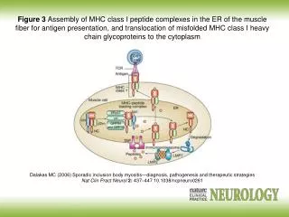
The binding of endogenous peptides occurs in the endoplasmic reticulum during biosynthesis of MHC gp I • After a chain a and b2mikroglobulin create in the ER, folding into the correct conformation and the mutual association and the association of an appropriate peptide, the complex is further processed in the Golgi apparatus and then is presented on the cell surface • Linked peptides are derived from proteins degraded by proteasome, proteasom degradate unneeded or damaged cytoplasmic proteins (labeled with ubiquitin), peptide fragments are transported into the ER by specific membrane pump TAP (transporter associated with antigen processing
Non-classical MHC gpI • HLA - E,-F,-G; CD1 molecules • Structurally similar to classical MHC gp • Are less polymorphic • There are only on some cells • They specialize in binding of specific ligands
HLA-E and HLA-G - occurs on the trophoblast cells • Complexes of HLA-E and HLA-G with peptides are recognized by inhibiting receptors of NK cells and contribute to the tolerance of the fetus in utero
MHC glycoproteins class II • The function of MHC gpII is the presentation of peptide fragments from protein whitch are ingested by cell on the cell surface so as to be recognized by T lymphocytes (helper, CD4) • Occur on the APC (dendritic cells, monocytes, macrophages, B lymphocytes) • 3 isotypes of MHC gpII (DR, DQ, DP)
MHC gp IIstructure • MHC gp II consist of 2 non-covalently associated transmembrane subunits a and b • The peptide binding site consists of N-terminal domains a1 and b1 • Binding of peptide is necessary for a stable MHC gp conformation and thus ensure its long presentation on the cell surface
MHC gp II peptide binding • MHC gpII bind peptides with a length of 15 to 35 aminoacides (but possibly longer - because the peptide binding site is open at both ends) • Certain MHC gp molecule binds peptides sharing common structural features - coupling motif • After a string a and b are created in ER, fold into the correct conformation and the mutual associated are connected with another transmembrane chain called invariant chain, which blocks the binding site for the peptide, this complex is further processed in the Golgi apparatus, secretory vesicles isolated from GA merge with endosomes, then split the invariant chain and peptide fragments from cell absorbed proteins bind into binding site of MHC gp and the complex is then presented on cell surface
Antigen presentation to T lymphocyte Signal:TCR– MHC gp I(II)+Ag peptid (APC) Co-stimulating signal: CD 28(T lymphocyte) – CD 80, CD 86 (APC)
MHC glycoproteinspolymorphism • HLA complex is located on chromosome 6 • For MHC gp is typical high polymorphism, there are up to hundreds of different forms of alelic isotypes (except the non-classical MHC gp, and DR a chain) • Codominant inheritance of alelic forms (Individualhas 3 cell surface isotypes of HLA molecules (HLA-A,-B,-C) mostly in 2 different alelic forms) • Polymorphism has a protective significance at individual and population level • MHC gp polymorphism causes complications in transplantation
HLA typing = determmination of HLA antigens on the surface of lymphocytesCarry out during the testing before transplantation and in determination of paternity • 1) Serotyping • Microlymfocytotoxic test • Allospecific serums (obtained from multiple natal to 6 weeks after birth, obtained by vaccination of volunteers, or commercially prepared sets of typing serums (monoclonal antibodies)) • Principle - the incubation of lymphocytes with typing serums in the presence of rabbit complement, then is added the vital dye which stained dead cells - cells carrying specific HLA are killed by cytotoxic Ab against the Ag, the percentage of dead cells is a measure of serum toxicity (forces and antileukocyte antibody titre) • Positive reaction is considered more than 10% dead cells (serological typing can be done also by flow cytometry
2) Molecular genetic methods • For typing are used hypervariable sections in the II. exon genes coding for HLA class II; to determine HLA class I is used polymorphism in II. and III. exon coding genes2a) PCR-SSP= Polymerase chain reaction with sequential specific primers • Extracted DNA is used as a substrate in a set of PCR reactions • Each PCR reaction contains primers pair specific for a certain allele (or group of alleles) • Positive and negative reactions are evaluated by electrophoresis, each combination of alleles has a specific electrophoretic painting
2b) PCR-SSO • PCR reaction with sequence-specific oligonucleotides Multiplication of hypervariable sections of genes coding HLA • Hybridization with enzyme or radiolabeled DNA probes specific for individual alleles 2c) PCR-SBT • Sequencing based typing • The most accurate method of HLA typing • We get the exact sequence of nucleotides, which compares with a database of known sequences of HLA alleles
T lymphocytes • cellular component of antigen-specific mechanisms • several different subsets of T lymphocytes • regulation of immune processes, the destruction of virus-infected cells or tumor cells • recognize antigen processed and presented by the APC • T cells are after activation stimulated to multiplication and differentiation into effector cells and part of them differentiate into the memory cells
T-lymphocytes development • T cells originate in bone marrow and then migrate to the thymus where they mature (abT lymphocytes), the final differentiation is after activation by antigen processed and presented by APC • gdT cells can develop outside the thymus (the minority population) Pluripotent hematopoietic stem cells Pro-thymocytes – double negative T cells - are coming from the bone marrow to the thymus, where they begin to rearrange TCRb genes, expressing on their surface, called pre-TCR (Composed of b chain, pre-TCRa and CD3 complex), then begin TCRa genes rearrangement Cortical thymocytes – double positive T cells - express on their surface TCR (composed of chains a, b and CD3) and CD4 and CD8 co-receptor (double positive T lymphocyte), at this stage occurs the selection of autoreactive cells and cells with dysfunctional TCR Medullary thymocytes (mature T cell) - retain the expression of CD4 or CD8, then migrate to secondary lymphoid organs
T-lymphocytes selection • Negative selection - the elimination of autoreactive cells, when thymocytes binds enough strongly by their TCR complex of MHCgp with normal peptides (from autoantigens)which are presented on surface of thymic cells thymocyte receives signals leading to apoptotic cell death PAE cells (peripherial antigen expressing cells) • Positive selection - the elimination of cells with dysfunctional TCR, positively are selected thymocytes that recognize MHC gp with low affinity, then maintain the expression of CD4 or CD8 (depending what class of MHC gp binds to the TCR). These mature T cells (Medullary thymocytes) leave the thymus and migrate to secondary lymphoid organs 98% of pro-thymocytes in the thymus during its development dies
T-lymphocytes surface markers • TCR - recognizes Ag peptide complexed with MHC gp • CD3 - TCR component, participation in signal transduction • CD4 or CD8 - co-receptors, binding to MHC gp • CD28 - costimulatory receptor, binds to CD80, CD86 on APC • CTLA-4 (CD152) - inhibitory receptor, binds to CD80, CD86
T-lymphocytes subpopulations • ab-T lymphocytes - have TCRab, major type (95%), thymus need in development, recognize antigens in the complex MHC-peptide gp • gd-T lymphocytes - (5%) may develop outside the thymus, some are able to recognize native Ag, apply in defense of the skin and mucous membranes
ab T-lymphocytes Expressing the CD4 co-receptor (co-receptor for MHC class II gp), precursors of helper T cells (TH), they can be classified according to the production of cytokines TH0 - produce a mixture of cytokines such as TH1 and TH2 TH1 - IL-2, IFNg (help macrophages ) TH2 - IL-4, IL-5, IL-6, IL-10 (B lymphocytes assistance) TH3 - TGFb Treg - regulatory T cells arise in the thymus from a part of autoreactive lymphocytes, suppress the activity of TH1 and partly function as TS, suppression of autoreactive T cell clones
ab T-lymphocytes Expressing the CD8 co-receptor (co-receptor for MHC gp class I), precursors of cytotoxic T cells (TC), or suppressor T cells (TS) TC - recognize cells infected by viruses or other intracellular parasites and some cancer cells TS - inhibit the function of other lymphocytes
TCR • TCR (T cell receptor) is heterodimer consisting of a and b (g,d) chain and associated CD3 complex,which is necessary for signal transfer (is connected with PTK) • "N-terminal parts of a and b (g,d) chain form the binding site for Ag
TCR development • The analogy with the formation of BCR • Chains b and d - correspond to IgH gene complex of immunoglobulins - V, D, J, C segments • Chains a and g- correspond to genes for L chains of immunoglobulins - V, J, C segments • Rearrangement of genes is similar to the BCR and performed by the same recombinases
TH1 immune response - inflammatory reaction • TH1 cells cooperate with macrophagesand transform them in activated (NO production - destroy intracellular parasites) • Activated macrophages secrete some cytokines (IL-1, TNF, ...) that help to stimulate T cells and stimulate local inflammation, which helps suppress infection • Interaction between TH1 cells and macrophages is a fundamental mechanism of delayed-type immunopathological reactions (DTH Delayed-type hypersensitivity)
The infected macrophage produces protein fragments derived from intracellular parasites, some of which are presented on the surface by MHC gp class II • Macrophages and dendritic cells stimulated by certain microorganisms produce IL-12 • TH precursor, which detects the infected macrophage and receives signals via the TCR, CD 28 and receptor for IL-12 and other adhesion and signaling molecules proliferates and differentiates to the effector TH1 cells that produce IFNg and IL-2. • IFNg activates macrophage NO synthase IL-2 is an autocrine growth factor for TH1 cells
TH2 immune response – help to B-lymphocytes • TH2 cells cooperate with B lymphocytes (which were stimulated by Ag) by cytokine production (IL-4, IL-5, IL-6) and direct intercellular contact • For stimulation of B lymphocytes is usually necessary cooperation between APC → TH2 cell → B lymphocyte • In minimal model, where the B cell becomes a good APC (CD80, CD86) is sufficient cooperation between TH2 cell → B lymphocyte
TH precursor, which detects the infected macrophage and receives signals through the TCR, CD 28 receptor for IL-4 receptor and IL-2 and other adhesion and signaling molecules proliferates and differentiates in the effector TH2, which provide B lymphocytes auxiliary signals via cytokines secreted by IL-4, IL-5, IL-6 and adhesion molecules through CD 40L, which bind to the costimulatory receptor on B lymphocytes CD 40 • Interaction between CD40 (B lymphocytes) and CD40L (TH2 cells) is essential for the initiation of somatic mutations, izotype switching and formation of memory cells • IL-4, IL-5, IL-6: stimulation of B lymphocytes
Assistance to B lymphocytes Specific direct assistance to B lymphocytes: • TH2 lymphocytes assisting B lymphocytes that were stimulated by the same Ag, which caused the rise of TH2 • To stimulate the secretion of cytokines by TH2 cell is sufficient signal via the TCR (signal through a costimulatory receptor CD28 is no longer necessary) • One clone of TH2 cells can provide specific assistance to B lymphocytes of different specificities (must present the relevant Ag peptides by MHC gp II, which are recognized by TCR)
Assistance to B lymphocytes Indirect assistance to B cells ("bystander help"): • TH2 lymphocytes assisting B lymphocytes that were stimulated by another Ag than that which caused the rise of TH2 • Contact between TH2 cell → B lymphocytes via adhesion molecules, cytokine secretion, binding CD40-CD40L • Danger of activation autoreactive B lymphocytes
Mutual regulation of activities TH1versus TH2 • Whether the TH precursor cell will develop into TH1 or TH2 decides cytokine ratio of IL-12 and IL-4 • IL-12 is produced by macrophages and dendritic cells stimulated by certain microorganisms • IL-4 is produced by activated basophils and mast cells • TH1 cytokines (mainly IFNg) inhibit the development of TH2 and stimulate the development of TH1 (IL-2 stimulates also TH2) • Cytokines produced by TH2 (IL-4, IL-10) inhibit the development of TH1 and stimulate the development of TH2 • TH3 development is stimulated by a specific cytokine environment (IL-4, IL-10, TGFb); TH3 produce TGFb and cooperate with B cells in MALT
Cytotoxic T lymphocytes stimulation • TC recognize cells infected with viruses or other intracellular parasites, and some tumor cells • Precursor of TC, which recognizes a complex of MHC gp I- antigenic peptide on the surface of APC via TCR and receives signals via CD 28 proliferates and differentiates to clone mature effector cytotoxic cells (CTL); TH1 cells help to TC by production IL-2 • Effector TC are spread by bloodstream into tissues; for activation of cytotoxic mechanisms is sufficient signal through the TCR (signal through a costimulatory receptor CD28 is no longer necessary)
Professional APC are dendritic cells or macrophages that are infected with virus, or swallowed antigens from dead infected, tumor or stressed cells • In order APC could activate the TC precursor, APC must be stimulated by contact with TH cells via CD 40, then the dendritic cell begins to express CD 80, CD86 and secrete cytokines (IL-1, IL-12) = change of resting APC in activated