1 / 8
80 likes | 82 Vues
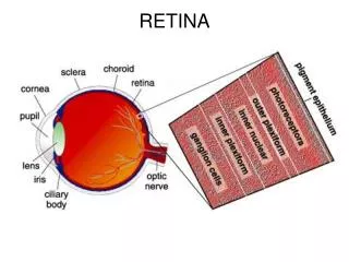
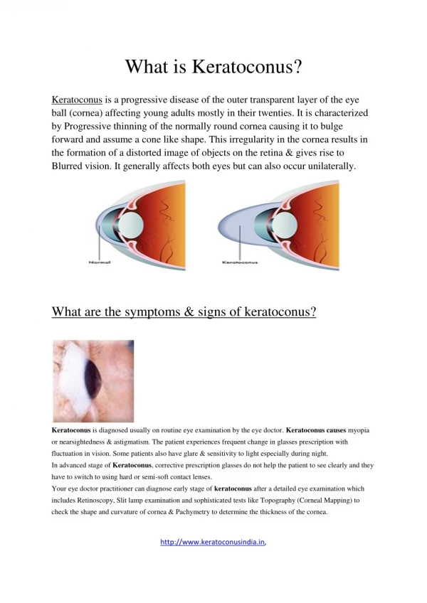
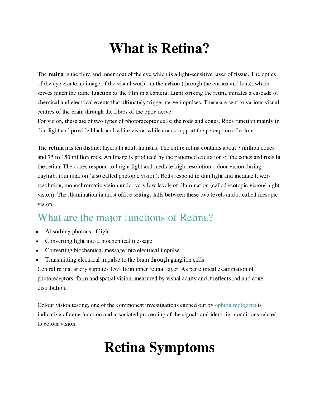
The retina is the third and inner coat of the eye which is a light-sensitive layer of tissue. The optics of the eye create an image of the visual world on the retina (through the cornea and lens), which serves much the same function as the film in a camera. Light striking the retina initiates a cascade of chemical and electrical events that ultimately trigger nerve impulses. These are sent to various visual centres of the brain through the fibres of the optic nerve.<br><br>For vision, these are of two types of photoreceptor cells: the rods and cones. Rods function mainly in dim light and provide black-and-white vision while cones support the perception of colour.

E N D
What is Retina? The retina is the third and inner coat of the eye which is a light-sensitive layer of tissue. The optics of the eye create an image of the visual world on the retina (through the cornea and lens), which serves much the same function as the film in a camera. Light striking the retina initiates a cascade of chemical and electrical events that ultimately trigger nerve impulses. These are sent to various visual centres of the brain through the fibres of the optic nerve. For vision, these are of two types of photoreceptor cells: the rods and cones. Rods function mainly in dim light and provide black-and-white vision while cones support the perception of colour. The retina has ten distinct layers In adult humans. The entire retina contains about 7 million cones and 75 to 150 million rods. An image is produced by the patterned excitation of the cones and rods in the retina. The cones respond to bright light and mediate high-resolution colour vision during daylight illumination (also called photopic vision). Rods respond to dim light and mediate lower- resolution, monochromatic vision under very low levels of illumination (called scotopic vision/ night vision). The illumination in most office settings falls between these two levels and is called mesopic vision. What are the major functions of Retina? Absorbing photons of light Converting light into a biochemical message Converting biochemical message into electrical impulse Transmitting electrical impulse to the brain through ganglion cells. Central retinal artery supplies 15% from inner retinal layer. As per clinical examination of photoreceptors; form and spatial vision, measured by visual acuity and it reflects rod and cone distribution. • • • • Colour vision testing, one of the commonest investigations carried out by ophthalmologists is indicative of cone function and associated processing of the signals and identifies conditions related to colour vision. Retina Symptoms
As far as the disease conditions resulting in retinal impairment are concerned, most instances of detachment start with a retinal tear. Here and there it precedes full separation. It typically has similar side effects. In the event that your retina gets torn, the liquid inside your eye can spill underneath and isolate the retina from its basic tissue. That is retinal separation. If you suspect there is a tear, go to the eye specialist. She can settle it in the workplace with a basic laser technique. On the off chance that you don't and it confines completely, you'll require more genuine surgery to repair it as s a part of an eye exam. The specialist will give you eye drops that extend your eye pupil (she'll call this widening your eyes). She'll utilize an extraordinary apparatus to investigate it and check whether your retina is confined. A retinal tear or another eye issue may cause: Floaters in your field of vision. Floaters are thick strands or bunches of strong vitreous gel that create as the gel ages and separates. Floaters frequently show up as dim spots, globs, strings, or specks. Floaters may likewise be caused by free blood or color from tears in the retina. Flashes of light or starts when you move your eyes or head. These are less demanding to see against a dull foundation. The concise flashes happen when the vitreous gel pulls on the retina (vitreous footing). These flashes typically show up at the edge of your visual field. Having floaters or flashes does not generally imply that you are going to have a retinal separation, however you ought not to overlook these side effects. Call your specialist to talk about whether you need an eye exam.
In the event that you have new or sudden flashes or floaters, haziness over piece of your visual field, or another loss of vision that does not leave, call your eye specialist or customary specialist immediately. Floaters and flashes might caution indications of retinal separation. A sudden shower of what seem, by all accounts, to be hundreds or thousands of minimal dark spots over the field of vision is a particular indication of blood or potentially shade in the vitreous gel and may demonstrate a retinal separation. This requires quick therapeutic consideration. In uncommon cases, a retinal separation can happen abruptly. The primary signs might be: A shadow or drape impact crosswise over piece of your visual field that does not leave. Since separations as a rule influence fringe (side) vision in the first place, you may not see an issue until the point that the separation has become greater. New or sudden vision misfortune. Vision misfortune caused by retinal separation has a tendency to deteriorate after some time. Sudden vision misfortune is a therapeutic crisis.
Retinal Disorders Common Symptoms of Retinal Disorders are Retinal detachment itself is painless. But warning signs almost always appear before it occurs or has advanced, such as: The sudden appearance of many floaters — tiny specks that seem to drift through your field of vision Flashes of light in one or both eyes Blurred vision Gradually reduced side (peripheral) vision A curtain-like shadow over your visual field Visual distortions, such as straight lines seeming bent Reduced central vision in one or both eyes Decreased intensity or brightness of colors A well-defined blurry spot or blind spot in your field of vision A general haziness in your overall vision • • • • • • • • • •
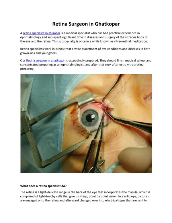
Retinal tear The retina is a layer of specialized nerve tissue lining the back of the eye that allows you to see. The inside of the eye is filled with a gel-like substance called the vitreous. A retinal tear occurs when there is an abnormal attachment between the vitreous gel and the retina. In this case the vitreous gel can tug on the retina causing it to tear. Retinal tears can develop at any age, but tend to occur more commonly in the elderly. Tears are also more common in people who have myopiawho have a history of previous retinal tear or a family history of retinal tears or detachments. The flashing lights are typically much more noticeable at night or in the dark and can sometimes become more intense with eye movement. Floaters, on the other hand, are usually much more noticeable in strong light. Some people have a lot of these symptoms while others notice hardly anything at all. Most retinal tears occur in the far peripheral retina, a part of the retina normally not used to see with. Therefore, most of the time there will be no noticeable change in the peripheral vision from the tear or the laser treatment. The laser treatment does not treat the floaters or flashing lights. These typically resolve gradually on their own over a period of weeks to months. Laser treatment around the tear is the treatment of choice to heal the Retinal break. Too Know More About Retina Disorder Retina Treatment And Surgery In Mumbai, India
Most retinal tears should be dealt with via fixing the retina to the back wall of the eye with laser surgery or cryotherapy(freezing treatment). Both of these systems make a scar that causes seal the retina to the back of the eye. This keeps liquid from going through the tear and under the retina, which ordinarily keeps the retina from segregating. These medicines cause practically no distress and might be performed in your ophthalmologist's office. How a laser surgery is performed? Laser surgery is done with laser, your ophthalmologist will use a laser to make little consumes around the retinal tear. The scarring that outcomes seals the retina to the hidden tissue, keeping a retinal separation. For freezing treatment, your eye specialist utilizes an extraordinary freezing test to apply serious freezing probe around the retinal tear. The outcome is a scar that secures the retina to the eye divider. Scleral clasp treatment includes setting an adaptable band (scleral clasp) around the eye to balance the power hauling the retina strange. The ophthalmologist frequently depletes the liquid under the isolates retina, enabling the retina to settle once more into its ordinary position against the back wall of the eye. This technique is performed in an operating room. The gas bubble pushes the retinal attack put against the back mass of the eye. Now and then this method should be possible in the ophthalmologist's office. After the surgery Your ophthalmologist will ask you to always keep up a specific set out position toward a few days. The gas air pocket will continuously vanish. What is Vitrectomy surgery? This is ordinarily used to settle a retinal separation and is performed in a working room. The vitreous gel, which is pulling on the retina, is expelled from the eye and for the most part supplanted with a gas bubble. Some of the time an oil bubble is utilized (rather than a gas rise) to keep the retina set up.
Your body own particular liquids will step by step supplant a gas bubble. An oil air pocket should be expelled from the eye at a later date with another surgical system. In some cases vitrectomy is joined with a scleral clasp. On the off chance that a gas bubble was set in your eye, your ophthalmologistmay prescribe that you keep your head in exceptional positions for a period. Try not to fly in a plane or go at high elevations until the point when you are told the gas bubble is no more. A quick increment in height can cause an unsafe ascent in eye weight. With an oil bubble, it is sheltered to fly on a plane. Most retinal separation surgeries (80 to 90 percent) are fruitful, despite the fact that a moment operation is at times required. Some retinal separations can't be settled. The advancement of scar tissue is the typical reason that a retina can't be settled. On the off chance that the retina can't be reattached, the eye will keep on losing sight and at last wind up noticeably visually impaired. After good surgery for retinal separation, vision may take numerous months to enhance and, now and again, may stay away for the indefinite future completely. Conditions We Treat Macular Degeneration Macular Hole Retinal Artery Occlusion Central Serous Retinopathy Retinoblastoma Endophthalmitis Cytomegalovirus (CMV) Retinal Infection Central Retinal Vein Occlusion Macular Pucker Hypertensive Retinopathy Retinal Hemorrhage Solar Retinopathy Retinitis Pigmentosa Retinal Tear Retinal Detachment • • • • • • • • • • • • • • •
Branch Retinal Vein Occlusion Intraocular Tumors Inherited Retinal Disorders Penetrating Ocular Trauma Pediatric and Neonatal Retinal Disorders Degenerative Myopia Diabetic Retinopathy Lattice Degeneration Uveitis Infectious Retinitis Macular Edema • • • • • • • • • • • We also Deal in Cataract, LASIK and keratoconus