Download
1 / 85
1.01k likes | 1.69k Vues
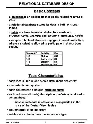
Spectroscopy, UV, IR and NMR

E N D
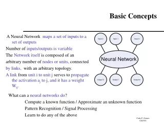
BASIC CONCEPTS OF ORGANIC SPECTROSCOPY Dr. Basavarajaiah S. M. M. Sc., Ph.D. Coordinator PG Department of Chemistry Vijaya college Bengaluru-56000.
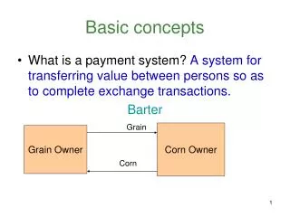
Spectroscopy Spectroscopy is a general term referring to the interactions of various types of electromagnetic radiation with matter. Exactly how the radiation interacts with matter is directly dependent on the energy of the radiation.
THE ELECTROMAGNETIC SPECTRUM Important: As the wavelength gets shorter, the energy of the radiation increases.
Electromagnetic radiation displays the properties of both particles and waves • The particle component is called a photon • The energy (E) component of a photon is proportional to the frequency . Where h is Planck’s constant and n is the frequency in Hertz (cycles per second) • E = hν • The term “photon” is implied to mean a small, massless particle that contains a small wave-packet of EM radiation/light – we will use this terminology in the course
Spectroscopy The higher energy ultraviolet and visible wavelengths affect the energy levels of the outer electrons. Infrared radiation is absorbed by matter resulting in rotation and/or vibration of molecules. Radio waves are used in nuclear magnetic Resonance and affect the spin of nuclei in a magnetic field.
UV-Vis Spectroscopy • Ultraviolet radiation stimulates molecular vibrations and electronic transitions. • Absorption spectroscopy from 160 nm to 780 nm. • Measurement absorption or transmittance. • Identification of inorganic and organic species.
UV/Vis Spectroscopy Visible (380-780 nanometers). Ultraviolet (UV) (10 – 380 nanometers). Below about 200 nm, air absorbs the UV light and instruments must be operated under a vacuum
1.Bathochromic Shift or Red shift: A shift of an absorption maximum towards longer wavelength (λ) or lower energy (E). • 2.Hypsochromic Shift or Blue Shift: A shift of an absorption maximum towards shorter wavelength (λ)or higher energy (E). • 3.Hyperchromic Effect: An effect that results in increased absorption intensity (ε). • 4.Hypochromic Effect: An effect that results in decreased absorption intensity (ε).
Wavelengths Absorbed by Functional Groups Again, demonstrates the moieties contributing to absorbance from 200-800 nm, because pi electron functions and atoms having no bonding valence shell electron pairs.
λmax = 455 nm λmax = 471 nm
INFRARED SPECTROSCOPY The IR region has lower energy than visible radiation and higher energy than microwave.
IR ABSORPTION BY MOLECULES • Molecules with covalent bonds may absorb IR radiation • Absorption is quantized • Molecules move to a higher energy state • IR radiation is sufficient enough to cause rotation and vibration • Radiation between 1 and 100 µm will cause excitation to higher vibrational states • Radiation higher than 100 µm will cause excitation to • higher rotational states
IR ABSORPTION BY MOLECULES • Absorption spectrum is composed of broad vibrational • Absorption bands • Molecules absorb radiation when a bond in the molecule vibrates • at the same frequency as the incident radiant energy • Molecules vibrate at higher amplitude after absorption • A molecule must have a change in dipole moment during • vibration in order to absorb IR radiation
IR ABSORPTION BY MOLECULES Absorption frequency depends on - Masses of atoms in the bonds - Geometry of the molecule - Strength of bond - Other contributing factors
DIPOLE MOMENT (µ) µ = Q x r Q = charge and r = distance between charges - Asymmetrical distribution of electrons in a bond renders the bond polar - A result of electronegativity difference - µ changes upon vibration due to changes in r - Change in µ with time is necessary for a molecule to absorb IR radiation
DIPOLE MOMENT (µ) • The repetitive changes in µ makes it possible for polar molecules • to absorb IR radiation • Symmetrical molecules do not absorb IR radiation since they • do not have dipole moment (O2, F2, H2, Cl2) • Diatomic molecules with dipole moment are IR-active • (HCl, HF, CO, HI) • Molecules with more than two atoms may or may not be • IR active depending on whether they have permanent • net dipole moment
Frequency Determination in IR • Excitation depends on atomic mass and how tightly they are bound • Hooke’s Law for 2 masses connected by a spring • C—H Bond: Reduced Mass = (12+1)/(12x1) = 13/12 = 1.08 • C—C Bond: Reduced Mass = (12+12)/(12x12) = 24/144 = 0.167 k = constant f (force constant) = bond strength m-term= µ= reduced mass
H H C C H H PRINCIPAL MODES OF VIBRATION Stretching Change in bond length resulting from change in interatomic distance (r) Two stretching modes - Symmetrical and asymmetrical stretching - Symmetrical stretching is IR-inactive (no change in µ) asymmetric symmetric
H H C C C C H H H H C C C C H H Bending - Change in bond angle or change in the position of a group of atoms with respect to the rest of the molecule Bending Modes - Scissoring and Rocking - In-plane bending modes (atoms remain in the same plane) - Wagging and Twisting Out-of-plane (oop) bending modes (atoms move out of plane) scissor rock twist wag in plane out of plane
Theoretical Vibrational Normal modes To locate a point in three-dimensional space requires three coordinates. To locate a molecule containing N atoms in three dimensions, 3N coordinates are required. The molecule is said to have 3N degrees of freedom. To describe the motion of such a molecule, translational, rotational, and vibrational motions must be considered. In a nonlinear molecule: 3 of these degrees are rotational and 3 are translational and the remaining correspond to fundamental vibrations; In a linear molecule: (Linear molecules cannot rotate about the bond axis) 2 degrees are rotational and 3 are translational. The net number of fundamental vibrations:
Vibrational modes of H2O (3 atoms –non linear) • Vibrational modes (degrees of freedom) = 3 x 3 - 6= 3 • These normal modes of vibration: • are a symmetric stretch, and asymmetric stretch, and a scissoring • (bending) mode.
Fundamental Vibrational modes of CO2 (3 atoms –Linear) • Fundamental Vibrational modes (degrees of freedom) = • 3 x 3 – 5 = 4 • These normal modes of vibration: • The asymmetrical stretch of CO2 gives a strong band in the IR at 2350 cm –1 (may noticed in samples due to presence of CO2in the atmosphere). • The two scissoring or bending vibrations are equivalent and therefore, have the same frequency and are said to be degenerate , appearing in an IR spectrum at 666 cm-1.
n-pentane 2850-2960 cm-1 sat’dC-H 3000 cm-1 1470 &1375 cm-1 CH3CH2CH2CH2CH3
cyclohexane no 1375 cm-1 no –CH3
1-decene unsat’d C-H 3020-3080 cm-1 910-920 & 990-1000 RCH=CH2 C=C 1640-1680
ethylbenzene 3000-3100 cm-1 Unsat’d C-H 1500 & 1600 Benzene ring 690-710, 730-770 mono-
o-xylene 735-770 ortho
styrene no sat’d C-H 1640 C=C 910-920 & 990-1000 RCH=CH2 mono
1-butanol 3200-3640 (b)O-H C-O 1o CH3CH2CH2CH2-OH
2-butanol O-H C-O 2o
tert-butyl alcohol O-H C-O 3o
methyl n-propyl ether no O--H C-O ether
2-butanone C=O ~1700 (s)