Download
1 / 12
130 likes | 299 Vues
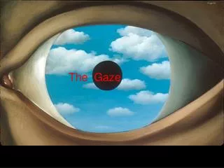
Sakina Dharsee Psychology - 260. Gaze Fixation and the Neural Circuitry of Face Processing in Autism. Introduction. Autism: Developmental disorder, unique profile of social and emotional behavior . Symptomatology : Diminished gaze fixation, lack of social or emotional reciprocity.

E N D
SakinaDharsee Psychology - 260 Gaze Fixation and the Neural Circuitry of Face Processing in Autism
Introduction • Autism: Developmental disorder, unique profile of social and emotional behavior. • Symptomatology: Diminished gaze fixation, lack of social or emotional reciprocity. • Previous research:Fusiformgyrus less activated in autistic patients. • Deficit:Which circuits are highly activated compared to normal?
Hypothesis • Fusiformgyrushypoactivation - Less time fixating on eye regions: Cause or consequence? • Hyperactivation in amygdala (region responsible for processing threatening, social and emotional cues)
Experimental Task Stimulation: Mock-up of MRI scanner. MRI Scan Eye movement: Eye-tracking device. • Study 1: • 14 autistic males (M:15.9, SD: 4.71) • 12 normal males (M: 17.1, SD: 2.78) • Task: Is the picture emotional or neutral? • Half straight, half turned. • Study 2: • 16 autistic males (M:14.5, SD: 4.60) • 16 normal males (M: 14.5, SD: 4.56) • Task: Is the picture familiar to you? • Ten photographs of family or friends.
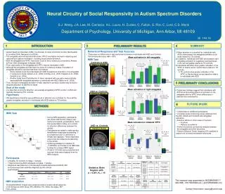
Result • Study 1: • Autistic group: • Longer judgement time for emotional and straight ahead faces. • Significantly less time fixating on eyes. • More activation in left amygdala. • Control group: • More activation in bilateral fusiformgyrus. a – Right fusiformgyrus b – Left fusiformgyrus
Study 2: • Control group: • More activation in bilateral fusiformgyrus. • Autistic group: • Significantly less time fixating on eyes. • More activation in right amygdala. • No differences in judgement time. Average duration of fixation on mouth, eye region and face in general.
Study 1: g – Left amygdala Study 2: e – Right amygdala
Discussion • Individuals with autism scan and process faces differently. • Deficits in processing emotional cues and in processing socially engaging faces. • Greater amygdala activation in autistic group for both studies. • Eye fixation creates a negative over-arousal mediated by the amygdala – diminished gaze fixation reduces this.
Negative over-arousal Increased sensitivity to social stimuli Diminished gaze fixation Hypoactivationof fusiformgyrus
Strengths and Limitations Limitations: • Strengths: • Experimental control. • Attempt to reduce performance anxiety. • Stimulation session. • Based solely on data obtained from males. • Inconsistency. • Future directions: • Lateralization of amygdala – left for emotion, right for familiarity? (Baas et al., 2004) • Treatment research: Can autistic patients be trained to scan and process faces normally, resulting in normal levels of fusiformgyrus activation?
Personal Opinion • Congruent with previous findings, and makes a new discovery. • Research can contribute towards the treatment and counselling of autistic children. • Leads into further research on the disorder – what are the circuits associated with other symptoms? Thank You! Any questions?