Download
1 / 1
10 likes | 105 Vues
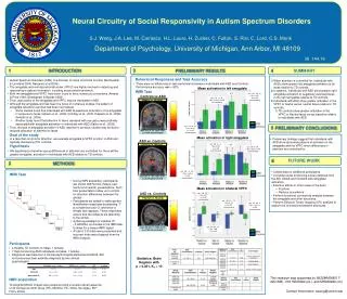
This study explores the activation patterns in the amygdala and ventral prefrontal cortex in individuals with Autism Spectrum Disorders (ASD) compared to typically developing controls. Preliminary results show differences in amygdala and VPFC activation with regard to attention and emotional stimuli processing. Future work includes examining social functioning correlations and connectivity analysis in other brain regions.

E N D
4 SUMMARY Happy vs. Neutral Fearful vs. Neutral Fearful vs. Neutral ASD vs. Controls ASD ASD ASD Controls Controls Controls Neural Circuitry of Social Responsivity in Autism Spectrum Disorders S.J. Weng, J.A. Lee, M. Carrasco, H.L. Louro, H. Zucker, C. Fulton, S. Risi, C. Lord, C.S. Monk Department of Psychology, University of Michigan, Ann Arbor, MI 48109 38 144.18 3 1 INTRODUCTION PRELIMINARY RESULTS Behavioral Responses and Task Accuracy • Autism Spectrum Disorders (ASD) is a disorder of social emotional function (Bachevalier & Loveland 2006; Nacewicz et al 2006). • The amygdala and ventral prefrontal cortex (VPFC) are highly involved in detecting and responding to salient information, including social-emotional stimuli. • Both the amygdala and VPFC have been found to have numerous connections (Amaral & Price 1984; Ghashghaei & Barbas 2002). • Thus, dysfunction of the amygdala and VPFC may be implicated in ASD. • Although the amygdala and has been the focus of numerous studies, the pattern of amygdala activation reported has been inconsistent. • Some studies found that individuals with ASD showed less activation in the amygdala in response to faces (Ashwin et al., 2006; Critchley et al., 2000; Dapretto et al., 2006; Gretlotti et al., 2005). • Another study found that attention to faces (assesed with eye gaze) was positively associated with amygdala activation in individuals with ASD (Dalton et al., 2005). • Thus, the lack of amygdala activation in ASD reported in previous studies may be due to reduced allocation of attention to faces. • When attention is controlled for, individuals with ASDs show greater left amygdala activation to all faces relative to TD controls. • In addition, individuals with ASD show greater right amygdala activation to negatively valenced faces in the right amygdala relative to TD controls. • Individuals with ASD show greater activation in the VPFC to fearful versus neutral faces relative to TD controls. • TD controls show greater activation in the VPFC to Neutral faces versus baseline relative to individuals with ASD. • There were no differences in task performance between individuals with ASD and Controls. Performance accuracy was > 92%. Mean activation in left amygdala fMRI Task N.S xyz: -20, -10, -8; t (9)= 2.92; p =0.08 (uncorrected) Controls vs ASD xyz: -14, -4, -8; t (9)= 2.03; p <0.05 (uncorrected) Contrast Vectors xyz: -14, -6, -8; t (9)= 3.32; p = 0.05 (uncorrected) 5 PRELIMINARY CONCLUSIONS xyz: -20, -4, -8; t (9)= 2.79; p <0.05 (uncorrected) Goal of the study • Preliminary findings suggest that individuals with ASD show abnormal patterns of activation in the amygdala and the VPFC when differences in attention are controlled for. Mean activation in right amygdala • In a task that controls for attention, we evaluate amygdala & VPFC function in ASD and typically developing (TD) controls. ASD vs. Controls xyz: 26, -8, -8; t (9)= 2.02; p <0.05 (uncorrected) N.S Hypothesis xyz: 28, 0, -26; t (9)=2.24; p<0.05 (uncorrected) • We hypothesize that when group differences in attention are controlled for, there will be greater amygdala activation in individuals with ASD relative to TD controls. Contrast Vectors FUTURE WORK 6 METHODS 2 • Collect data on additional participants • Correlate social functioning scores obtained from the ADI, ADOS and Vineland with amygdala activation. • Examine effects in other areas of the brain: • Fusiform • Nucleus accumbens • Perform functional connectivity analysis between • the amygdala and other structures. • Perform Diffusion Tensor Imaging (DTI) analysis to explore the connections between structures. xyz: 36 , -2, -20; t (9)= 3.43; p <0.05 (uncorrected) fMRI Task xyz: 28, -2, -26; t (9)= 2.27; p <0.05 (uncorrected) • During fMRI acquisition, participants are shown 250ms face (happy, sad, fearful and neutral) presentations. Such brief presentations allow us to control for attention differences between the groups. • Participants are asked to make gender identification responses by pressing “1” to a male face and “2” whenever a female face appears. These responses ensure that the subjects are attending to the stimuli. • A jittering paradigm of variable ITI ( 0-6000ms) is included in the fMRI task to allow for a clearer fMRI signal. • A total of 120 trials were presented and incorrect trials were dropped from the fMRI analysis. Mean activation in bilateral VPFC xyz: -46, 32, 2; t (9)= 3.27; p <0.05 (uncorrected) xyz: -36, 14, -12; t (9)= 6.13; p <0.001 (uncorrected) xyz: -42, 46, 2; t (9)= 1.87; p <0.05 (uncorrected) Contrast Vectors xyz: 34, 34, -14; t (9)= 2.98; P= 0.08 xyz: -46, 34, 2 ; t (9)= 3.27; p <0.05 (uncorrected) Participants • 4 Healthy TD controls (3 males, 1 female) • 7 High-functioning ASD individuals (6 males, 1 female) • Diagnoses was based on a clinical psychologists administered ADOS, ADI • and consensus best estimate diagnosis by two clinical • psychologists. Statistics: Brain Regions with p < 0.001, Ke > 10 This research was supported by 5K22MH068017-02(CSM), U19 HD35482 (CL), and MH066496 (CL) fMRI acquisition T2 weighted BOLD images were acquired using a reverse spiral sequence of 40 contiguous 3mm slices (TR= 2000ms; TE= 30ms, flip angle= 80o; FOV= 22cm). Contact Information: sweng@umich.edu