
Functional Gene Networks in Genome Activity: Co-Expression Analysis
Explore co-expression networks across different functional states of genome activity to understand gene relationships and complex diseases. Compare varied cell types and stimulation experiments to identify functional gene networks through correlation analysis. Our research aims to develop algorithms and software for medical research focusing on gene expression profiles, cytometric profiles, and biomarker identification for inflammatory diseases. Contact us to learn more about our work and collaborations.

Functional Gene Networks in Genome Activity: Co-Expression Analysis
E N D
Presentation Transcript
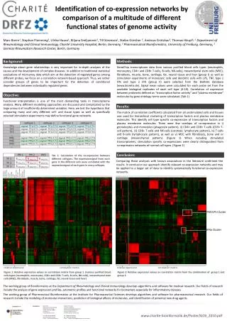
Identification of co-expression networks by comparison of a multitude of different functional states of genome activity Marc Bonin1, Stephan Flemming2, Ulrike Haase1, Biljana Smiljanovic3, Till Sörensen1, Stefan Günther 2, Andreas Grützkau3, Thomas Häupl1: 1 Department of Rheumatology and Clinical Immunology, Charité UniversityHospital, Berlin, Germany, 2Pharmaceutical Bioinformatics, University of Freiburg, Germany, 3 German Rheumatism Research Center, Berlin, Germany Background: Methods: Knowledge about gene relationships is very important for in-depth analyses of the causes and the development of complex diseases. In addition to traditional statistical evaluations of microarray data which aim at the detection of regulated genes among different probes, we focus on a correlation network-based approach. Thus, we rather consider groups of genes to be important for the detection of conditional dependencies between individually regulated genes. GeneChip transcriptome data from various purified blood cells types (neutrophils, monocytes, CD4+ and CD8+ T-cells, B-cells, NK-cells), mesenchymal stem cells (MSC), fibroblasts, muscle, bone, cartilage, fat, neural tissue and liver (group I) as well as stimulation experiments of monocytic cells and dendritic cells with LPS, TNF, type 1 IFN and type 2 IFN (group II) were selected from the BioRetis database (www.bioretis.de). Signal mean values were calculated for each probe set from the available biological replicates of each cell type (4-10). Correlation of expression between probesets defined as "transcription factor activity" and "plasma membrane" molecules by gene ontology terms were calculated. (Tab 1) Objective: Functional interpretation is one of the most demanding tasks in transcriptome analysis. Many different modelling approaches are discussed and complicated by the large amount of insufficiently determined variables. Here, we test the hypothesis that comparing many and very different cell- and tissue types as well as specifically selected stimulation experiments may define functional gene networks. Results: The matrix of correlation coefficents calculated from all unstimulated cells and tissues was used for hierarchical clustering of transcription factors and plasma membrane molecules. This identify cell type specific co-expression of transcription factors and plasma membrane molecules. There were few overlaps of co-expression in i) gronolozytes and monozytes (phagocyte pattern), ii) CD4+ and CD8+ T-cells (CD3+ T-cell pattern), iii) CD8+ T-cells and NK-cells (cytotoxic lymphocyte pattern), iv) T-cells and B-cells (lymphocyte pattern), as well as v) MSC with fibroblasts, bone and or cartilage (mesenchymal pattern). (Figure 1) When including stimulated transcriptomes, stimulation specific co-expressions were clearly distinguished from co-expression networks of normal cell types. (Figure 2) Conclusion: Tab 1: Calculation of the co-expression between different celltypes. The expressionsignal from each gene in the different cells were correlated with the expressionsignal of each gene in every celltypes. Comparing these analyses with known associations in the literature confirmed the results. In conclusion our approach identify relevant co-expression networks and may be applied to a larger set of data to identify systematically functional co-expression networks. 1 fibroblasts 2 monozytes 3 CD4+ T-cells 4 muscle 5 bone 6 cartilage 7 MSC 8 fat 9 CD8+ T-cells 10 B-cells 11 neural tissue 12 liver 13 NK-cells 14 neutrophils TNF/LPS Cluster IFNa Cluster relative expression correlation matrix relative expression correlation matrix Figure 1: Relative expression values an correlation matrix from group 1. (various purified blood cells types (neutrophils, monocytes, CD4+ and CD8+ T-cells, B-cells, NK-cells), mesenchymal stem cells (MSC), fibroblasts, muscle, bone, cartilage, fat, neural tissue and liver) Figure 2: Relative expression values an correlation matrix from the combination of group 1 and group 2. The working group of Bioinformatics at the Department of Rheumatology and Clinical Immunology develops algorithms and software for medical research. Our fields of research include the analysis of gene expression profiles, zytometric profiles and functional networks for biomarkers especiallyfor inflammatory diseases. The working group of Pharmceutical Bioinformatics at the Institute for Pharmaceutical Sciences develops algorithms and software for pharmaceutical research. Our fields of research include the modeling of molecular interactions, prediction of biological effects of molecules, and identification of potential new drug agents. Contacts: Marc Bonin Department of Rheumatology and Clinical Immunology Charité University Hospital Charitéplatz 1 D-10117 Berlin Germany Tel: +49(0) 30 450 564 056 Fax: +49(0) 30 450 513 957 E-Mail: marc.bonin@charite.de Stephan Flemming University of FreiburgInstitute of Pharmaceutical SciencesPharmaceutical BioinformaticsHermann-Herder-Strasse 9D-79104 Freiburg i. Br. Germany Tel: +49(0) 761-203-4873 Fax: +49(0) 761-203-6366 E-Mail: stephan.flemming@pharmazie.uni-freiburg.de www.charite-bioinformatik.de/Poster/GCB_2010.pdf