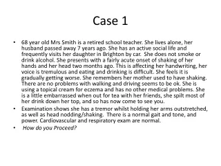
Case 1
Case 1. 82 y.o. female Retrosternal burning Long standing dysphagia. Upper esophagus. Larynx. Upper esophagus. Lower esophagus. Diverticulum Duplication of esophagus Congenital double lumen esophagus Fistula None of the above. Case 2. 49 y.o. woman

Case 1
E N D
Presentation Transcript
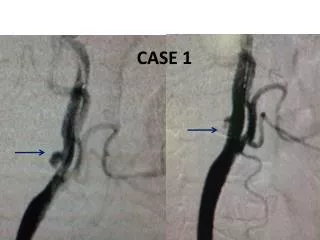
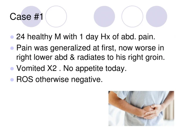
Case 1 • 82 y.o. female • Retrosternal burning • Long standing dysphagia
Larynx Upper esophagus Lower esophagus
Diverticulum • Duplication of esophagus • Congenital double lumen esophagus • Fistula • None of the above
Case 2 • 49 y.o. woman • Chronic idiopathic pancreatitis • Severe pain – on narcotics • ERCP X 2: failed cannulation of Wirsung due to severe stricture
EUS Calcifications Head Wirsung 11mm
EUS – guided pancreaticogastrostomy Francois et al. Gastrointestinal Endoscopy 2002 • 4 patients • 3 of 4 had satisfactory pain relief • 1 year follow-up
Case 3 • 51 y.o. woman with mild RUQ pain • No relevant medical history • No medications • Normal physical examination
CT PVP Late phase Hepatic artery phase
MRI T1Contr hepatic artery phase
Differential diagnosis • Atypical hemangioma • Hemangiosarcoma • Necrotic tumor • Peliosis hepatis • Other
Etiology • Wasting illness • Carcinomatosis • Drugs (anabolics, steroids, oral contraceptives, azathioprine) • Immunosuppressed patients • AIDS – bacillary angiomatosis peliosis • Idiopathic
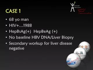
Case 4 • 51 y.o. man • Diabetes mellitus type II, target organ damage, no prior abdominal operations • Abdominal pain, vomiting, weight loss • Palpable mass at the right upper quadrant
Differential diagnosis- Partial pseudoobstruction- Retroperitoneal fibrosis- Sclerosing peritonitis- Paraduodenal hernia- Amyloidosis
Cocoon syndrome • First described in 1978 • Idiopathic , sclerosing , encapsulating peritonitis • - congenital - idiopathic - secondary (peritoneal dialysis, peritoneovenous shunts, beta-blockers, peritoneal TB, GI malignancy)