Download
1 / 41
440 likes | 747 Vues
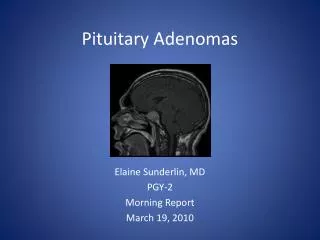
Investigation protocols in pituitary adenomas functional and non functional. Introduction. Pituitary gland – Pituitary fossa • Mass: 5 gms • DIMENSIONS – 7mm (Ht) – 9mm (AP) – 11m(transverse) originates from Rathke’s pouch and infundibulum. Introduction. 15% of intracranial tumors

E N D
Investigation protocols in pituitary adenomasfunctional and non functional
Introduction Pituitary gland – Pituitary fossa • Mass: 5 gms • DIMENSIONS – 7mm (Ht) – 9mm (AP) – 11m(transverse) originates from Rathke’s pouch and infundibulum
Introduction • 15% of intracranial tumors • Present as incidental finding in 5-20% • Broadly divided (a) functional (b) non functional
INVESTIGATION PROTOCOL • History and physical examination • Neuro- ophthalmology: Acuity, field, fundus and movements • Hormone levels Basal hormone and dynamic testing Aim- hypersecretory state or insufficiency • Radiology (a) X-Rays (b) MRI (c) NCCT/CECT • Routine blood investigation
Presentation • Mass effect • Hyper secretion/ hypo secretion • Incidental finding • Apoplexy
Complete history and physical examination • Eyes – visual acuity, visual field, fundoscopy • Neck- thyroid ,carotid bruit • Chest-gynaecomastia, galactorrhea • Abdomen-striae, obesity • Extremities-edema, enlargement • Skin-pigment, hair, bruises
MASS EFFECT • Visual disturbances – Visual field defect usually very insidious and slowly progressive – Diplopia – Visual acuity • Hydrocephalus • Headache • Cranial nerve palsies • Raised intracranial pressure
Apoplexy Acute presentation secondary to tumour haemorrhagic necrosis – Headache – Vomiting – Blindness – Ocular paresis – Altered level of consciousness
Prolactin Galactorrhoea , amennorrhoea, osteoporosis • G.H Acromegaly, organomegaly, D.M, • ACTH Cushing’s disease, Diabetes mellitus, osteoporosis, obesity, hypertension • TSH Hyperthyroidism, cardiac dysrhythmia, heat intolerance
Radiology • X- Rays: Widening of sella Destruction of sellar floor Relation of median sphenoidal septum Aeration of sphenoid sinus- conchal sclerotic mixed
NCCT+ CECT head/ sella with thin coronal cuts: findings as seen in X-Rays iso dense to adjacent brain parenchyma intense contrast enhancement calcifications uncommon (< 5%) apoplexy- hyper density
MRI brain: Sagittal T!WI and coronal T1WI sellar and parasellar region with/without contrast 2.5mm thin contiguous slices and 5mm slices axial T2WI of whole brain. Normal pituitary is iso intense to gray matter on T1WI with contrast enhancing Pituitary adenoma classified based on size: microadenoma <10mm macroadenoma >10mm giant pit adenoma >40mm
Macro adenoma – they are hypo to isointense to gray matter on T1WI, contrast enhancing • Micro adenomas- Dynamic contrast study done by 5 T1WI turbo spin 3mm thin slices repetitively at 20,40,60,80,100 sec after 10ml contrast injection at 2ml/sec. • Micro adenoma enhance and wash out quickly as compared to normal gland post contrast and hence appear hypointense • deviation of stalk • bulging of inferior and superior margin
Hardy classification • Pituitary adenoma: Grade 0- size < 10 mm, sella normal Grade 1- size < 10 mm, sella expanded Grade 2- size > 10 mm , sella expanded Grade 3- size > 10 mm, focal Destruction Grade 4- size > 10 mm, diffuse destruction Grade 5- distant spread
Based on extension • Suprasellar 0- none A- supra sellar cistern B- ant recess of third ventricle obliterated C- floor of third ventricle grossly displaced • Parasellar D- intracranial (intradural) E- into or beneath the cavernous sinus
Screening studies for pituitary lesion • Hormone excess serum prolactin serum IGF-1 serum LH, FSH serum A sub unit serum TSH urinary24 hr cortisol • Hormone deficiency serum cortisol serum T4, free T3 serum testosterone (men) serum estradiol (women)
Dynamic test to identify pituitary hypersecretion Dynamic stimulation/suppression testing may be useful in select cases to further evaluate pituitary reserve and/or for pituitary hyperfunction • Acromegaly Oral glucose test- • Cushings syndrome/disease- (a)low dose dexamethasone (b)low dose dexamethasone +CRH (c)high dose dexamethasone (d)Inferior petrous sampling + CRH
Dynamic test to identify pituitary deficiency • ACTH – low dose ACTH by giving 1 mcg iv and S. cortisol after 30 min less than 18 mcg/dl identifies central adrenal deficiency
Prolactinoma • 30 to 50% of endocrine active tumors • Clinical features: • Amennorhoea infertility, loss of libido, oligospermia • Galactorrhoea in 80% females and 30% men • Majority are microadenomas • 30% of them in women are self limiting
Prolactinoma • Prolactin < 25 ng/ ml normal 25- 150ng/ml prolactinoma, stalk effect, drugs , Hypothyroid > 150ng/ml- prolactinoma • Hook effect- even large elevations will show normal PRL levels on testing due to large size of molecules. Do serial dilutions
Cushings disease • 15% of all pituitary adenomas in adults • 90% microadenomas • Common in women • 55 % pit adenoma in children • Clinical features: Central obesity, purple striae, hypertension, diabetes, ecchymosis, poor wound healing, lipid abnormalities, neuropsyhiatric problems
Cushings disease • Best screening test-24 hr UFC level 95-100% sensitivity, 400 mcg/day of UFC is diagnostic. • midnight plasma cortisol of 5.2mcg/dl is diagnostic of cushings • Low dose dexamethasone test- 1 mg of dexa at 11.00 am and measurement of s. cortisol at 8.00 am <5 mcg/dl- normal 5-10 mcg/dl equivocal >10mcg diagnostic • Plasma corticotropin level- >20pg/ml diagnostic >10 pg/ml suggestive <5pg/ml corticotroph independent
Cushings disease • High dose dexa suppression test- if corticotrops >10 pg/ml . 2mg of dexa given every 6hrly for 2 days, if > 69% fall in 24 hr UFC (pre and post dexa ) is 100% specific for CD • 8 mg dexamethasone test -8 mg dexamethasone is given at 11.00 pm and drop in >50% s. cortisol indicates CD • Corticotropin releasing hormone stimulation test- I mcg/kg CRH iv in morning, if increases >35% corticotropin level at 15, 30 min above baseline yields 100% specificity and 93% sensitivity for CD
Cushings disease • Inferior petrosal sinus sampling • classical clinical and biochemical CD features with MRI negative patient • equivocal suppression and stimulation test Diagnostic accuracy is 80-100% , blood samples are obtained at basal and 3,5,10 min after CRH administration and ips/ps ratio calculated ips/ps >3 CD ips/ps <2 ectopic rarely 2-3 ectopic IPS gradient helps in lateralization of adenoma
Acromegaly • 4th decade of life • 10.7 years • Constitute 20% of all pituitary tumors • Preop duration 10 years to diagnosis in adults and 3.1 years in children • Pleuri hormonal
ACROMEGALY • Prepuberty-gigantism & precocious puberty • Pubescent-amenorrhea, hypogonadism • Adults-skeletal and soft tissue overgrowth and deformities, cardiac ,neuromuscular, respiratory, endocrine, metabolic complications and neoplastic transformation
Random GH – not useful gives false positive and false negative results • Insulin like growth factor 1 (IGF-1) – best for screening represents average daily GH secretion • Oral glucose GH suppression testing – gold standard to confirm diagnosis :75 mg of glucose load normally suppresses GH > 2ng/ml RIA. GH nadir >2ng/ml RIA with adenoma confirms it • GHRH stimulation test
ACROMEGALY • Chest and abdomen imaging for ectopic GHRH secreting tumors • Empty sella shows pituitary infarction • Scintigraphy • Ancillary tests Blood glucose, urine, cardiac and respiratory Screening for colorectal neoplasia
Clinically Acromegaly, MRI pit adenoma, GH>5ng/ml If GH<5ng/ml IGF-1, elevated If no Oral glucose suppression test confirms it rarely MRI negative , measure GHRH levels, CECT abd /chest
Thyrotroph adenoma • TSH secreting tumors • 1-2% of pit adenomas • Mixed hormonal secretion- 30% GH, PRL, Gonadotropins • 90% macroadenomas • Mean duration pt 9 yrs • Clinical features of goitre, warm skin, heat intolerance, cardiac arrhythmias and other hyperthyroid features,
Thyrotroph adenoma • Lab investigations TSH, Free t4,t3by direct method a-subunit, PRL, GH, SHB Iodine scan/USG of thyroid Dynamic testing with TRH
Clinical suspicion, MRI –pit adenoma, baseline TSH, free T4/T3,a-sub unit,PRL,GH TSH normal, a-sub unit/TSH ratio <5.7 in normogonads,<29.1 in hypergonad, TSH elevated<0.7 in normogonads, <1.0 in hypergonads MRI equivocal, TRH stimulation test
Gonadotropinomas • 7-15% of pit adenomas • 40-50 % macroadenomas secrete gonadotropins • Clinical features of mass effect: visual symptoms, hypogonadism, amennorrhea, hypothyroid, hypocortisolism
Gonadotropinomas • Lab investigations basal hormonal levels TRH stimulated gonadotropins, and sub units normally causes absent FSH response and no more than 33% increase in LH and b- LH primary hypogonadism LH,FSH elevated and don’t respond to TRH gonadotropinomas have greater than 60% increase in b-LH levels
Multidisciplinary approach Hormonal status-endocrinologist Visual field –orthoptist Monitor tumor recurrence –radiologist Clinical observation-neurosurgeon Blood test-biochemist