CASE PRESENTation ON SPINE FRACTURE
480 likes | 957 Vues
CASE PRESENTation ON SPINE FRACTURE. PRESENTED BY SHERIN JOHN. PHYSICAL ASSESMENT General Appearance The patient is 25 yrs of age ,male . He is conscious and oriented, with the following vital signs; BP :110/70mm of Hg PR :92bpm

CASE PRESENTation ON SPINE FRACTURE
E N D
Presentation Transcript
CASE PRESENTationON SPINE FRACTURE PRESENTED BY SHERIN JOHN
PHYSICAL ASSESMENT • General Appearance • The patient is 25 yrs of age ,male . • He is conscious and oriented, with the following vital signs; • BP :110/70mm of Hg • PR :92bpm • RR :24cpm • Temp :36.80 • SPO2 :96%
DEMOGRAPHIC DATA • NAME : Mr. M F A • AGE/ SEX : 25 years /Male MR NO :192406 • DATE OF ADMISSION :18/12/2012 • DIAGNOSIS :Fracture D7 • SURGERY :Posterior Fixation Of D6 – D8 ON 18/12/2012
Skin • Fair complexion • Abrations present • Head • Cut wound over scalp and swelling present • Consciousness and Orientation • Awake and conscious and GCS is 15/15 on admission. • Oriented to persons(knows some of his relatives) • Place (he can tell where he is) • Time(knows the day,date,and always asking time.
Eyes • Right eye is normal • Left eye lid hematoma present • Pupils equally round and reactive to light • Conjunctiva,cornea and lens normal • Ears • No discharges found • Hearing normal • Nose • Pink nasal mucosa • No unusual nasal discharges • No tenderness in sinuses • Mouth • Pink and moist oral mucosa and free of swelling and lesions
Neck and Throat • No palpable lymph nodes • No masses and lesions seen • Chest and Lungs • Pain present in right lower chest and breathing difficulty • After investigations;ct chest and x-ray chest shows haemopneumothorax with rib fracture and right side ICD inserted • Cardio Vascular • ECG reports showsclear and no changes noted
Gastro Intestinal • No Tender Ness of Abdomen and it is soft. • GenitoUrinary • With Foley catheter fr.18 • Musculoskeletal • Unable to move his left arm • Has pain during Examination • Cannot perform ADL • Numbness over both lower limbs • Neurologic • Patient is mentally alert and oriented with circumstances. • Able to follow Commands • GCS is 15/15 • Right radial nurve injury.
PATIENT HISTORY • Past Medical History • There is no history of surgeries done • Present Medical History • Patient brought to ER by Red crescent c/o difficulty in breathing and pain in the right lower chest and back pain and numbness in both lower limbs due to RTA on 18-12-12 and patient was admitted in icu and he underwent posterior spine fixation of D6-D8 on the same day
VITAL SIGNS • BP :110/70mm of Hg • PR :92bpm • RR :24cpm • Temp :36.80 • SPO2 : 96% • INVESTIGATIONS DONE FOR THE PATIENT • CT scan Brain without contrast • CT scan chest and abdomen with contrast • CT scan cervical spine,thoracic spine and lumbar spine. • Blood Investigations • TREATMENT • Surgical Intervention: Posterior spine fixation of D6-D8
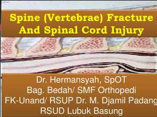
Spine AnatomyThe Five Segments of the Spine • The spinal column is one of the primary support structures in the human skeleton. our spine is made up of a column of vertically stacked bones, called vertebrae, which surround and protect the spinal cord. There are 33 separate vertebrae that are divided into five segments: cervical, thoracic, lumbar, sacral and coccyx. Each of the vertebra lines up much like blocks on top of one other. There are discs between each vertebra, which act as shock absorbers between each bony vertebra. The vertebrae, while interconnected, operate relatively independently, which gives your back a degree of flexibility while enabling it to provide a tremendous amount of support.
The Nervous System Running down the spine, and protected by the vertebra, are some of the most important components of the human body's nervous system. In particular, the spinal cord is protected by an opening in the back of the vertebrae called the foramen. Other nerves extend from the spinal cord through spaces between each vertebra. The Disc In between the vertebrae are the discs. The discs act as a cushion between the vertebral bones. Each disc consists of a soft jelly-like center and a tough outer material that holds the disc's shape and position in the spine.
The Facets On the back of the vertebrae are two sets of facets; one set at the top of the vertebra (called the "superior facets") and one set at the bottom (called the "inferior facets"). The superior facets face up and make contact with the downward facing inferior facets on the next vertebra forming the facet joint. The facet joints are the points at which two vertebrae contact each other. The facet joints act as a hinge on which two vertebrae move when bending forward and backward.
The Ligaments, Tendons and Muscles • A variety of soft tissue holds the entire spinal column in place and allows it to flex. Ligaments and tendons are fibrous bands of tissue that attach to bone. Ligaments connect two or more vertebrae and help stabilize the spine. Tendons attach muscle to bone allowing the spine to move when the muscles contract and relax. • Spinal Blood Supply • The function of the vascular system is to nourish each cell in the body. This includes the vertebral column, spinal cord, neural elements, muscles, and other related structures. Blood and Its ImportanceBlood contains plasma (fluid), red blood cells (erythrocytes), white bloods cells, and platelets.
Vascular system of the spine Red = ArteryBlue = Vein 1 Carotid Artery2 Aortic Arch3 Thoracic Aorta4 Abdominal Aorta5 Iliac Artery6 Internal Jugular Vein7 Superior Vena Cava8 Inferior Vena Cava9 Iliac Vein
Spinal Cord and Nerve RootsThe spinal cord originates in the brain, exiting through a hole at the skull base called the foramen magnum and coursing through the spinal canal of the cervical, thoracic and upper lumbar spine before ending most commonly between the first and second lumbar vertebrae.Nerve roots exiting from the lower end of the spinal cord continue as a structure called the caudaequina, or horse's tail, to provide nerves to the lower trunk, legs, bowels, bladder and sexual organs.
Spinal Cord and Nerve Roots • The spinal cord originates in the brain, exiting through a hole at the skull base called the foramen magnum and coursing through the spinal canal of the cervical, thoracic and upper lumbar spine before ending most commonly between the first and second lumbar vertebrae. • Nerve roots exiting from the lower end of the spinal cord continue as a structure called the caudaequina, or horse's tail, to provide nerves to the lower trunk, legs, bowels, bladder and sexual organs. Damage to the nerves can cause pain, tingling, numbness or weakness in the area where the nerve travels. Damage to the spinal cord at any level can cause many symptoms, from paralysis to numbness.
Functions of the Spine • The three main functions of the spine are to: • Protect the spinal cord, nerve roots and several of the body’s internal organs. • Provide structural support and balance to maintain an upright posture. • Enable flexible motion. • Regions of the Spine • Typically, the spine is divided into four main regions: cervical, thoracic, lumbar and sacral. Each region has specific characteristics and functions. • Cervical Spine • The neck region of the spine is known as the Cervical Spine. This region consists of seven vertebrae, which are abbreviated C1 through C7 (top to bottom). These vertebrae protect the brain stem and the spinal cord, support the skull, and allow for a wide range of head movement.
The first cervical vertebra (C1) is called the Atlas. The Atlas is ring-shaped and it supports the skull. C2 is called the Axis. It is circular in shape with a blunt peg-like structure (called the Odontoid Process or “dens”) that projects upward into the ring of the Atlas. Together, the Atlas and Axis enable the head to rotate and turn. The other cervical vertebrae (C3 through C7) are shaped like boxes with small spinous processes (finger-like projections) that extend from the back of the vertebrae. • Thoracic Spine • Beneath the last cervical vertebra are the 12 vertebrae of the Thoracic Spine. These are abbreviated T1 through T12 (top to bottom). T1 is the smallest and T12 is the largest thoracic vertebra. The thoracic vertebrae are larger than the cervical bones and have longer spinous processes. In addition to longer spinous processes, rib attachments add to the thoracic spine’s strength. These structures make the thoracic spine more stable than the cervical or lumbar regions. In addition, the rib cage and ligament systems limit the thoracic spine’s range of motion and protect many vital organs.
Lumbar Spine • The Lumbar Spine has 5 vertebrae abbreviated L1 through L5 (largest). The size and shape of each lumbar vertebra is designed to carry most of the body’s weight. Each structural element of a lumbar vertebra is bigger, wider and broader than similar components in the cervical and thoracic regions. • The lumbar spine has more range of motion than the thoracic spine, but less than the cervical spine. The lumbar facet joints allow for significant flexion and extension movement but limit rotation. • Sacral Spine • The Sacrum is located behind the pelvis. Five bones (abbreviated S1 through S5) fused into a triangular shape, form the sacrum. The sacrum fits between the two hipbones connecting the spine to the pelvis. The last lumbar vertebra (L5) articulates (moves) with the sacrum. • Immediately below the sacrum are five additional bones, fused together to form the Coccyx (tailbone).
The Pelvis and the Skull • Although not typically viewed as part of the spine, the pelvis and the skull are anatomic structures that closely inter-relate with the spine, and have a significant impact on the patient’s balance. • Spinal Curves • When viewed from the front (Coronal Plane) the healthy spine is straight. (A sideways curve in the spine is known as scoliosis.) When viewed from the side (Sagittal Plane) the mature spine has four distinct curves. These curves are described as being either kyphotic or lordotic. • A kyphotic curve is a convex curve in the spine (i.e. convexity towards the back of the spine). The curves in the thoracic and sacral spine are kyphotic. • A lordotic curve is concave (i.e. concavity towards the back of the spine), and is found in the cervical and lumbar levels of the spine.
Vertebral StructuresAll vertebrae consist of the same basic elements, with the exception of the first two cervical vertebrae. The outer shell of a vertebra is made of cortical bone. This type of bone is dense, solid and strong. Inside each vertebra is cancellous bone, which is weaker than cortical bone and consists of loosely knit structures that look somewhat like a honeycomb. Bone marrow, which forms red blood cells and some types of white blood cells, is found within the cavities of cancellous bone.
Vertebrae consist of the following common elements:Veterbral BodyThe largest part of a vertebra. If looked at from above it generally has a somewhat oval shape. When looked at from the side, the vertebral body is shaped like an hourglass, being thicker at the ends and thinner in the middle. The body is covered with strong cortical bone, with cancellous bone within.
Pedicles • These are two short processes, made of strong cortical bone, that protrude from the back of the vertebral body. • Laminae • Two relatively flat plates of bone that extend from the pedicles on either side and join in the midline. • Processes • There are three types of processes: articular, transverse and spinous. The processes serve as connection points for ligaments and tendons.
Endplates • The top (superior) and bottom (inferior) of each vertebral body is “coated” with an endplate. Endplates are complex structures that “blend” into the intervertebral disc and help support the disc. • Intervertebral Foramen • The pedicles have a small notch on their upper surface and a deep notch on their bottom surface. When the vertebrae are stacked on top of each other the pedicle notches form an area called the intervertebral foramen. This area is of critical importance as the nerve roots exit from the spinal cord through this area to the rest of the body.
Facet Joints • The joints in the spinal column are located posterior to the vertebral body (on the backside). These joints help the spine to bend, twist, and extend in different directions. Although these joints enable movement, they also restrict excessive movement such as hyperextension and hyper-flexion (i.e. whiplash).
Each vertebra has two facet joints. The superior articular facet faces upward and works like a hinge with the inferior articular facet (below). • Like other joints in the body, each facet joint is surrounded by a capsule of connective tissue and produces synovial fluid to nourish and lubricate the joint. The surfaces of the joint are coated with cartilage that helps each joint to move (articulate) smoothly. • Intervertebral Discs • Between each vertebral body is a "cushion" called an intervertebral disc. Each disc absorbs the stress and shock the body incurs during movement and prevents the vertebrae from grinding against one another. The intervertebral discs are the largest structures in the body without a vascular supply. Through osmosis, each disc absorbs needed nutrients. • Each disc is made up of two parts: the annulus fibrosis and the nucleus pulposus.
Annulus Fibrosus • The annulus is a sturdy tire-like structure that encases a gel-like center, the nucleus pulposus. The annulus enhances the spine’s rotational stability and helps to resist compressive stress. • The annulus consists of water and layers of sturdy elastic collagen fibers. The fibers are oriented at different angles horizontally similar to the construction of a radial tire. Collagen gains its strength from strong fibrous bundles of protein that are linked together. • Nucleus Pulposus • The center portion of each intervertebral disc is a filled with a gel-like elastic substance. Together with the annulus fibrosus, the nucleus pulposus transmits stress and weight from vertebra to vertebra. • Like the annulus fibrosus, the nucleus pulposus consists of water, collagen and proteoglycans. However, the proportion of these substances in the nucleus pulposus is different. The nucleus contains more water than the annulus.
Types of Spinal Fractures • A compression fracture of the lumbar (lower) spine.
There are different types of spinal fractures. Doctors classify fractures of the thoracic and lumbar spine based upon pattern of injury and whether there is a spinal cord injury. Classifying the fracture patterns can help to determine the proper treatment. The three major types of spine fracture patterns are flexion, extension, and rotation. • Flexion Fracture Pattern • Compression fracture. While the front (anterior) of the vertebra breaks and loses height, the back (posterior) part of it does not. This type of fracture is usually stable and rarely associated with neurologic problems.
Axial burst fracture. The vertebra loses height on both the front and back sides. It is often caused by a fall from a height and landing on the feet. • Extension Fracture Pattern • Flexion/distraction (Chance) fracture. The vertebra is literally pulled apart (distraction). This can happen in accidents such as a head-on car crash, in which the upper body is thrown forward while the pelvis is stabilized by a lap seat belt. • Rotation Fracture Pattern • Transverse process fracture. This fracture is uncommon and results from rotation or extreme sideways (lateral) bending, and usually does not affect stability.
Fracture-dislocation. This is an unstable injury involving bone and/or soft tissue in which a vertebra may move off an adjacent vertebra (displaced). These injuries frequently cause serious spinal cord compression A side-view of a fracture-dislocation of a thoracic vertebra. A magnetic resonance imaging (MRI) scan of a fracture-dislocation in the thoracic spine. Note the disruption of the spinal cord.
Surgical treatment for thoracic spine fracture-dislocation without neurological deficit. The surgical treatment depends on the type of spinal injury and the level .
Posterior spine fixation with screw and rods and bone grafting • Posterior spine fixation with screw ,rod and cage and bone grafting • Posterior and anterior decompression and stabilization with implants
The transitional anatomy of the thoracolumbar spine makes it vulnerable to injury from high-energy vehicular crashes and falls. The definitive management of patients with thoracolumbar spinal fractures is dependent on the presence and extent of neurologic injury, the presence and magnitude of acute deformity, and an estimate concerning spinal stability. It is well established that neurologic deficits generally improve without surgery. Nonsurgical treatment leads to decreased pain and improved function. Although there is a dearth of high-quality studies comparing surgical with nonsurgical treatment, the natural course of thoracolumbar fractures usually is benign, and nonsurgical methods should be the standard treatment with few exceptions.
Complications of spine fracture • The most serious complication of vertebral fracture is spinal cord injury which can result in paralysis. • Infection • Nerve damage in displaced fracture. • Faulty alignment of healdvertibre(malunion) • Gastro intestinal disease • Chronic pain • Complications of spine surgery • Ophthalmic complication associated with prone positioning in spine surgery • Post operative spinal wound infections • Various thromboembolism in spine surgery
PRIORITIZATION OF NURSING PROBLEMS • Impaired physical mobility related to neuro muscular impairment and vertebral body fracture. • Anxiety related to perceived effects of injury on life style and unknown fracture. • Bowel incontinence: reflexes related to lack of voluntary sphincter control secondary to spinal cord injury.
NURSING HEALTH TEACHING • Encourage early ambulation. • Encourage him to perform passive and active excercises. • Instruct patient in methods of safe ambulation----brace and wheel chair etc. • Discuss prevention of recurrent fractures. • Teach symptoms needing attention such as numbness,decreasedfunction,increasedpain,elevated temperature. • Teach the importance of follow up care. • Encourage follow up medical supervision to monitor for union problems. • Encourage adequate balanced diet to promote bone and soft tissue healing. Conclusion • A case of RTA patient with fracture of D7 and partially able to move his both lower extremity. • Surgical treatment posterior fixation of D6-D8done. • Patient is able to move his legs slowly • Health education given on physiotherapy and bowel training